接第一篇:
3. 2 CMN inhibited the tumor growth of xenograft mice
To investigate the active anti-lung cancer fractions of CM,xenograft mouse models were constructed, and the experimental scheme was presented in Fig. 2A. The tumor volume was significantly decreased post-treatment, notably in CM and CMN groups (P < 0. 05, Fig. 2B). Additionally, the tumor weights decreased significantly in CM and CMN groups (Fig. 2C). Immunohistochemistry experiments showed that CM and CMN significantly reduced Ki67 protein expression in tumor tissue sections (Fig. 2D). Hematoxylin and eosin (H&E) staining of tumor tissues also demonstrated a comparable trend, as depicted in Fig. 2E. Extensive tumor cell necrosis was observed in the tumor pathological sections from the CM and CMN groups, indicating that CM and CMN exerted a potent destructive impact on tumor tissue. Mice were weighed every two days during the entire administration phase. The differences in body weight among the blank, model, and each active fraction of CM groups were insignificant. Nevertheless, in the DDP and CM groups, body weight decreased over time and differed substantially from that of the model group (P < 0. 01, Fig. 2F). Our preliminary studies demonstrated that CM intervention markedly suppressed tumor growth (P < 0. 05) at the dosage of 1 g·kg-1[9]. The relatively high concentration of CM administration was associated with marked body weight reduction in experimental mice within this therapeutically effective dose range. In contrast, mice in the CMN group did not lose weight after administration of the drug. The results showed that CMN exerted a significant antitumor effect without losing weight compared to CM.
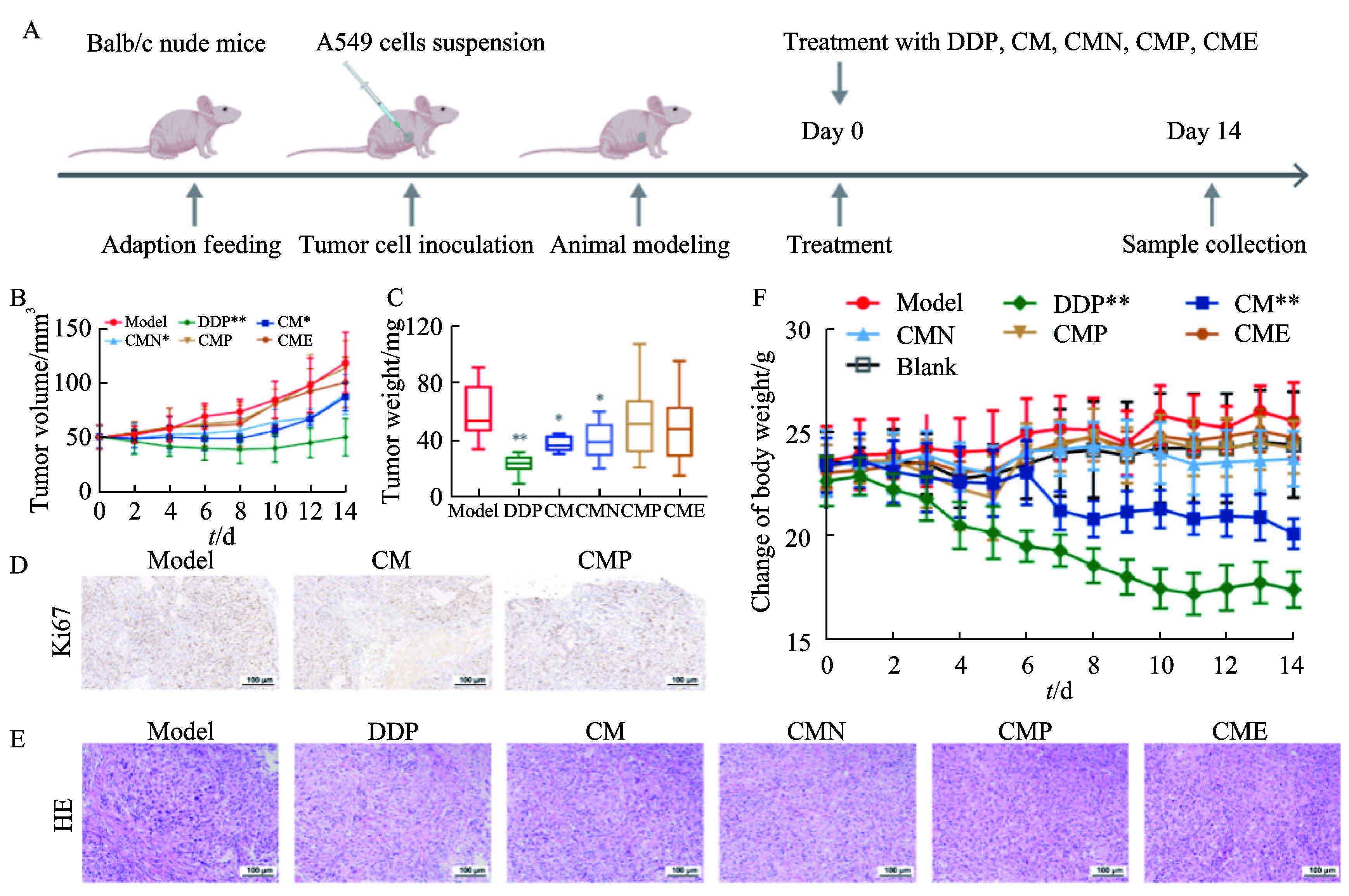
Note: A. Schematic flow of the pharmacological assay; B. Growth curves of the tumor volume; C. Tumor weights in xenograft mice; D. Histopathological section micrographs of the tumor tissues( IHC staining, 200×); E. Micrographs of tumor histopathological sections( H&E staining, 200×);F. Variations in nude mouse body weight. Data were presented as xˉ±s, n = 8. *P<0. 05, **P<0. 01 vs. the model group.
Fig. 2 CMN inhibited lung cancer progression in xenograft mice
3. 3 Metabolomic analysis
3. 3. 1 Metabolomic analysis of serum samples
CMN was observed to exert significant inhibitory effects on the tumor growth of xenograft mice based on animal experiments. The effect of CMN on serum differential metabolites in xenograft mice was examined via untargeted metabolomics. The total ion chromatograms exhibited well-formed peaks (Fig. S1A). Through comparison with MS-DIAL and NIST databases, 97 metabolites were identified, predominantly comprising amino acids, glycerides, unsaturated fatty acids, and carbohydrates. Clear separation between the CMN group and the model group was effectively demonstrated via Principal Component Analysis (PCA)and partial least-squares discrimination analysis (PLS-DA)(Fig. 3A). To validate the model quality, 200 response permutation tests were conducted. Model parameters from the permutation analysis displayed an upward trend, suggesting that the OPLS-DA model was stable with accurate predictions(Fig. 3B). A total of 38 metabolites were altered after CMN administration compared to the model (Table S2). The volcano plot indicated that 19 differential metabolites had increased and 19 had decreased (Fig. 3C). Differential metabolites in serum are shown in the heat-maps (Fig. 3D). These results suggested that tumor xenotransplantation triggered alterations in endogenous metabolites in nude mice, with CMN reversing such changes. The distinction between the CMN and model groups suggested that CMN exerted a significant regulatory influence on abnormal metabolite levels in xenograft mice. It is proposed that CMN might exert its anti-lung cancer effect by modulating the levels of these metabolites. The 38 differential metabolites were entered into the Metaboanalys 5. 0 online analysis website for processing. The 11 differential metabolite pathways were identified from the model and CMN groups with P < 0. 05 (Fig. 3E),including the TCA cycle, alanine, aspartate and glutamate metabolism, glyoxylate and dicarboxylate metabolism, and arginine biosynthesis. Serum metabolomics results indicated that CMN could influence the changes of metabolites through the regulation of these metabolite pathways, thereby exerting antilung cancer effects.
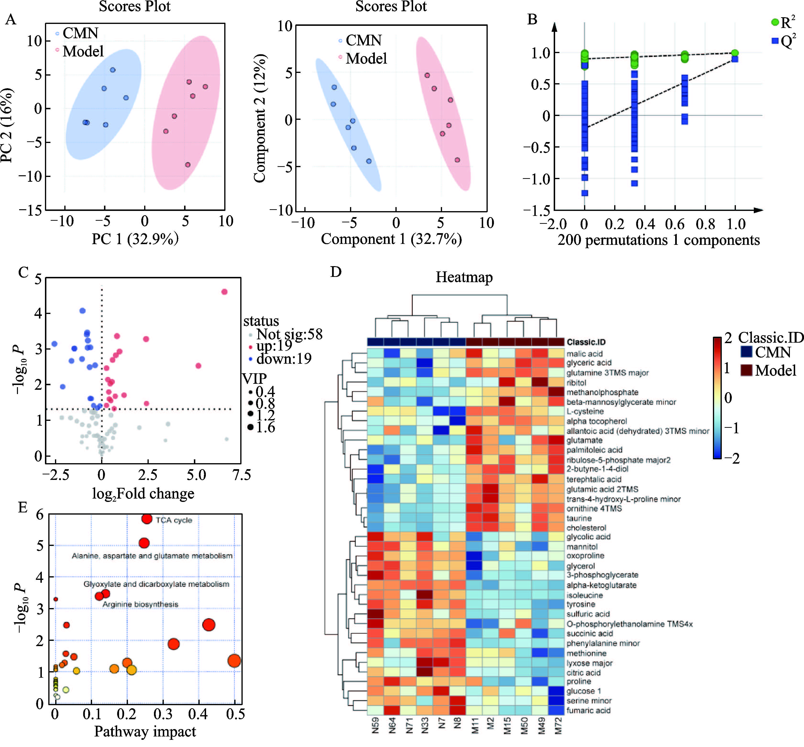
Note: A. PCA and PLS-DA score scatter plots of serum metabolites; B. Permutation tests of OPLS-DA models; C. Volcanic plot of differential metabolites; D. Heatmap displaying differentially expressed metabolites between model and CMN groups. Upregulated metabolites were shown as red dots, while downregulated ones were shown in blue; E. KEGG enrichment analyses for metabolic pathways in serum samples
Fig. 3 The mechanism of CMN treating lung cancer was explored by serum metabolomics
3. 3. 2 Metabolomic analysis of tumor tissues
To further validate the serum metabolome results and investigate the metabolic influence of CMN on tumors, we analyzed metabolites in tumor samples from xenograft mice using metabolomic analysis. Total ion chromatograms showed well-formed peaks (Fig. S1B). Additionally, the clear separation among different groups was revealed via PCA and PLS-DA score plots (Fig. 4A). The results of the 200-response replacement test indicated that the model was stable and the predictions were accurate (Fig. 4B). These findings suggested that xenotransplantation of tumors could induce changes in endogenous metabolites in nude mice. A total of 102 metabolites were identified using MS-DIAL and NIST database comparisons, mainly including compounds such as amino acids, fatty acids, glycerides, and carbohydrates. The volcano plot showed that 3 differential metabolites had increased and 41 had decreased (Fig. 4C). The differential metabolites in the tumors were displayed through the heat maps (Fig. 4D).Relative to the model group, 44 metabolic biomarkers underwent changes following CMN treatment (Table S3). These findings indicated that CMN might exert its anti-lung cancer activity by adjusting the levels of these metabolites. The differential metabolites were entered into the Metaboanalys 5. 0 online analysis site for processing to obtain metabolic pathways. The potential pathways associated with the anti-lung cancer effect of CMN were identified with P < 0. 05 (Fig. 4E). Findings primarily highlighted involvement in valine, leucine and isoleucine biosynthesis, one carbon pool by folate, glyoxylate and dicarboxylate metabolism, glycine, serine and threonine metabolism, purine metabolism, arginine biosynthesis, butanoate metabolism,pantothenate and coA biosynthesis, and TCA cycle. In conclusion, the metabolic pathways that played a role together in tumor metabolomics and serum metabolomics were one carbon pool by folate, glyoxylate and dicarboxylate metabolism, arginine biosynthesis, butanoate metabolism, TCA cycle pathway. The results showed that CMN mainly regulated the above pathways and thus exerted anti-lung cancer effects.
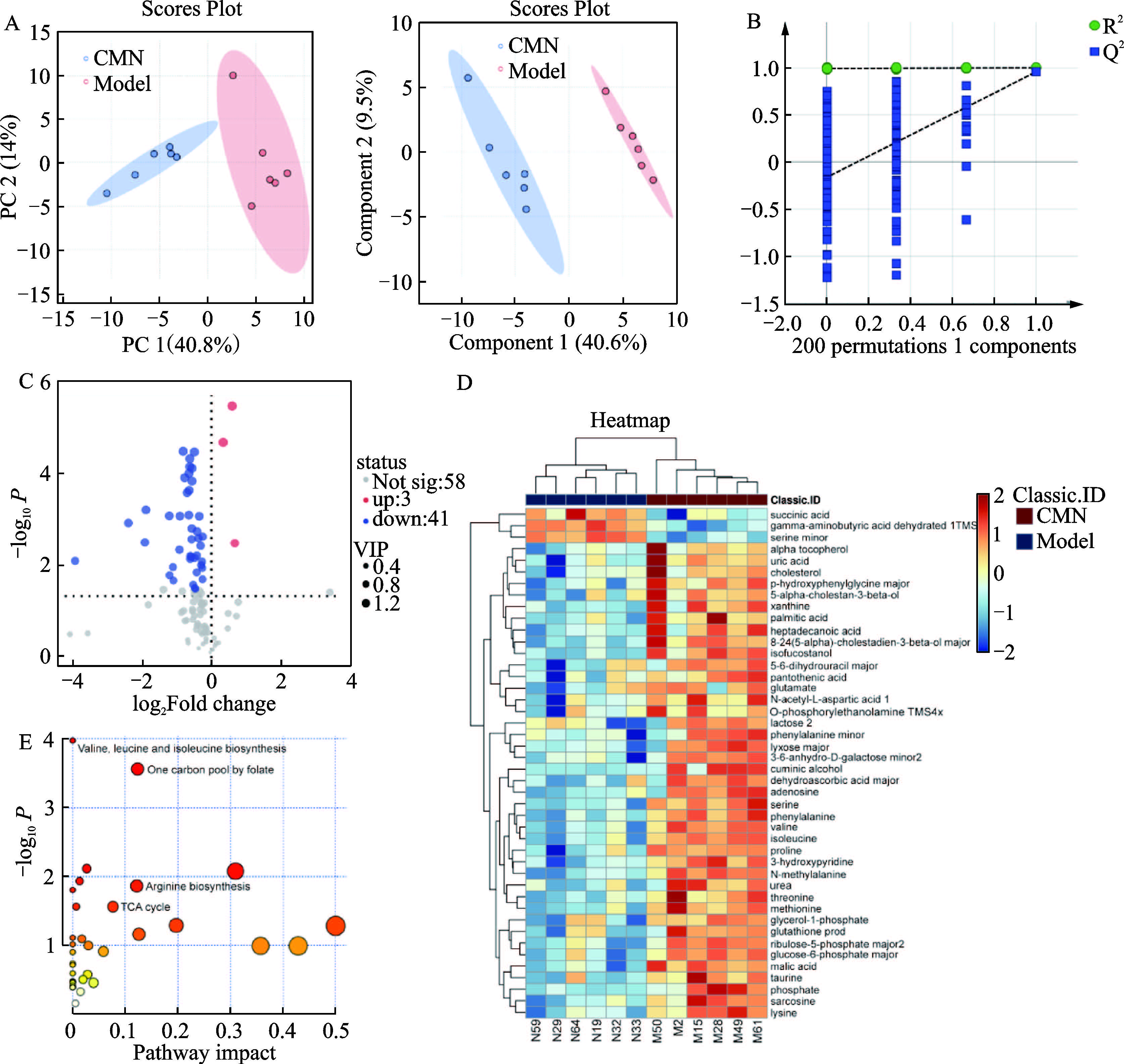
Note: A. PCA and PLS-DA score scatter plots of tumor metabolites; B. Replacement test for OPLS-DA model; C. Volcanic plot of differential metabolites; D. Heatmap displaying differentially expressed metabolites between model and CMN groups;E. KEGG enrichment analysis for metabolic pathways in tumor samples.
Fig. 4 The mechanism of CMN treating lung cancer was explored by tumor metabolomics
3. 4 CMN inhibited the proliferation of lung cancer cells
The results of animal experiments demonstrated that CMN exhibited a significant inhibitory effect on tumor growth in xenograft mice, with minimal side effects. To further elucidate its anti-tumor mechanisms, a series of tumor cell assays focusing on lung cancer cells were conducted. Initially, tumor cells were exposed to CMN at multiple concentrations for 24, 48, and 72 h. The results revealed that the survival rate of tumor cells declined in a concentration-dependent pattern. Specifically,the IC50 values of CMN for tumor cells were 10. 020 μg·mL-1 at 24 h, 1. 822 μg·mL-1 at 48 h, and 2. 417 μg·mL-1 at 72 h(Fig. 5A). These results suggested that CMN markedly suppressed tumor cell proliferation in a concentration-dependent pattern. To assess the ability of CMN to inhibit tumor cell proliferation and migration, its effects on colony formation were investigated. Relative to the control group, colony numbers declined notably as CMN concentration increased (Fig. 5B). Quantitative analysis further confirmed that CMN inhibited colony formation in a dose-dependent manner (Fig. 5C). The number of colonies and the relative proliferation survival rate were negatively correlated with the concentration of CMN administered. Wound healing assays were used to assess the effect of CMN on the migration ability of lung cancer cells. In the CMN group,the rate of wound closure was significantly diminished, with the inhibitory effect intensifying in a concentration-dependent manner( Fig. 5D). These results indicated that CMN effectively suppressed lung cancer cell migration in a concentration-dependent pattern (Fig. 5E). To explore whether CMN exerts cell cycleblocking effects, the changes in cell cycle distribution of tumor cells following treatment with CMN (2, 4, and 8 μg·mL-1) for 24 hours were analyzed through flow cytometry. The results indicated that CMN treatment significantly increased the proportion of tumor cells in the G1 phase while simultaneously reducing the percentage of cells in the S phase (Fig. 5F). This suggested that CMN arrested lung cancer cells in the pre-G1 phase,thereby inhibiting DNA synthesis, reducing the transition of lung cancer cells from diploid to tetraploid, and ultimately suppressing cell proliferation.
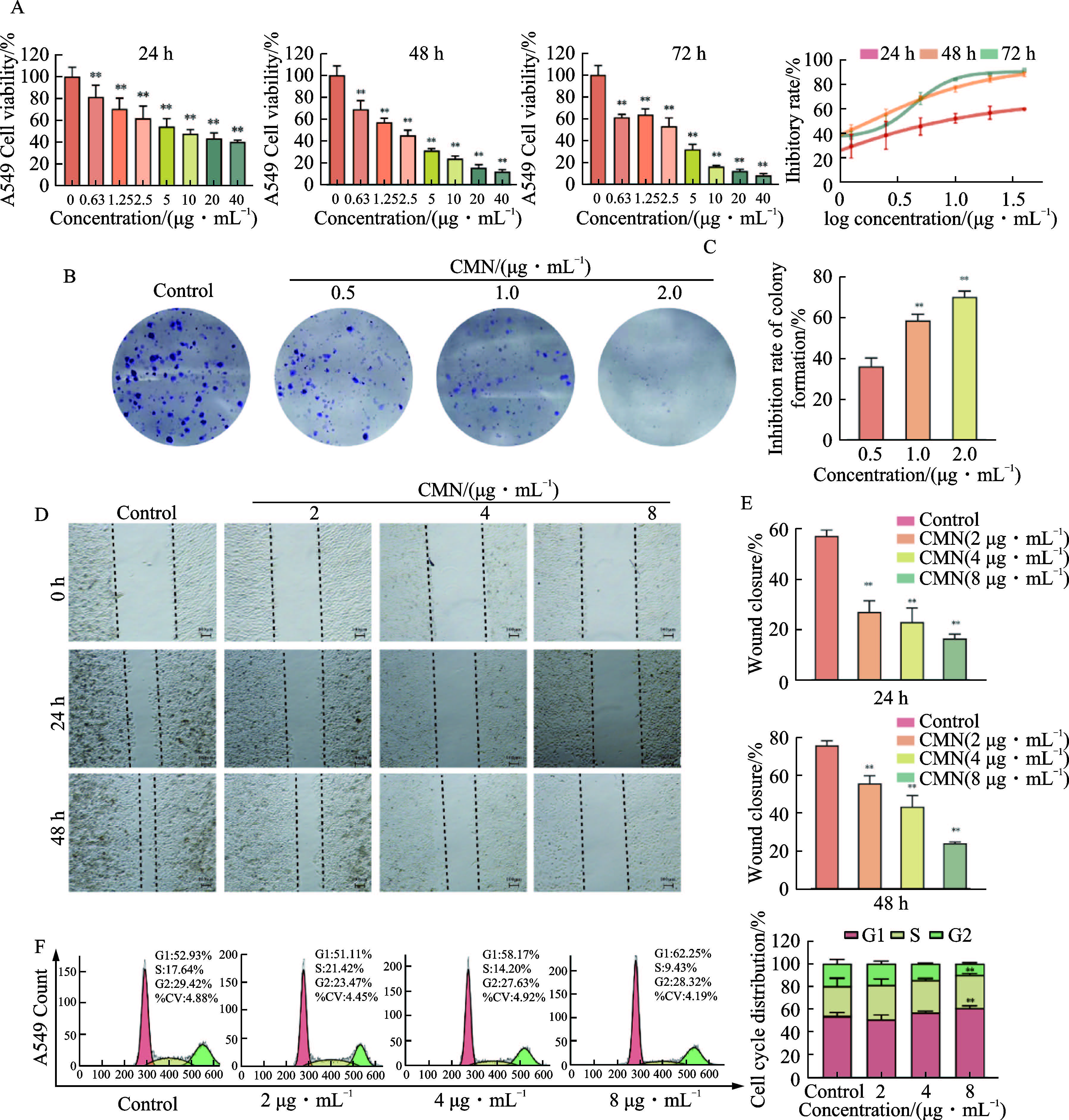
Note: A. Influence of CMN on the proliferation of tumor cells; B. Influence of CMN on colony formation of tumor cells. ; C. Quantitative analysis of the colony-forming ability of tumor cells by CMN; D. Influence of CMN on the cell migration of tumor cells; E. Quantitative analysis of CMN on the migration of tumor cells; F. Influence of CMN on the cell cycle of tumor cells. Data were shown as xˉ±s, **P< 0. 01 vs. the control group.
Fig. 5 CMN inhibited the proliferation and migration of lung cancer cells
3. 5 Metabolomic analysis of cell samples
The metabolic pathways of CMN treatment for lung cancer were explored using serum and tumor metabolomes in the previous part, and we wanted to validate the above results through tumor cell metabolomic studies in this part of the study. Tumor cell samples from the control, CMN-5 (5 μg·mL-1 CMN), and CMN-10 (10 μg·mL-1 CMN) groups were subjected to metabolomic analysis using GC-MS. The method suitability of GC-MS analysis was validated by normalizing the ionic strength of the base peak to 100% (Fig. S1C). PCA and PLS-DA score plots revealed distinct separations between the control, CMN-5, and CMN-10 groups (Fig. 6A). Through comparison with MSDIAL and NIST databases, 96 metabolites were identified, primarily comprising amino acids, fatty acids, glycerides, and carbohydrates. Screening of differential metabolites was performed with criteria of VIP ≥ 1 and P < 0. 05. Compared to the control group, 49 metabolite levels were significantly altered in the CMN-5 group (Table S4), while 47 metabolite levels were significantly changed in the CMN-10 group (Table S5). Volcano plots indicated that CMN-5 treatment led to 4 metabolites had increased and 45 having decreased, whereas CMN-10 treatment induced 5 metabolites having increased and 42 having decreased (Fig. 6B). Clustering heatmap analysis showed that the CMN-5 and CMN-10 groups clustered together, distinct from the Control group (Fig. 6C), highlighting the significant metabolic regulation by CMN treatment. These differential metabolites were further analyzed using the Metaboanalys 5. 0 online platform, with pathways enriched based on a P < 0. 05 threshold. Eight differential metabolite pathways were identified, including galactose metabolism, TCA cycle, alanine, aspartate and glutamate metabolism, arginine biosynthesis, phenylalanine, tyrosine and tryptophan biosynthesis, phenylalanine metabolism, butanoate metabolism, starch and sucrose metabolism(Fig. 6D). The identified differential metabolites are intricately linked to the anti-tumor mechanisms of CMN based on these findings. A comparative analysis of the metabolic pathways from serum, tumor, and cellular metabolomes revealed a convergence on the TCA cycle pathway. Collectively, these results suggested that CMN exerted its anti-cancer effects by modulating metabolite changes through the inhibition of TCA cycle pathway.
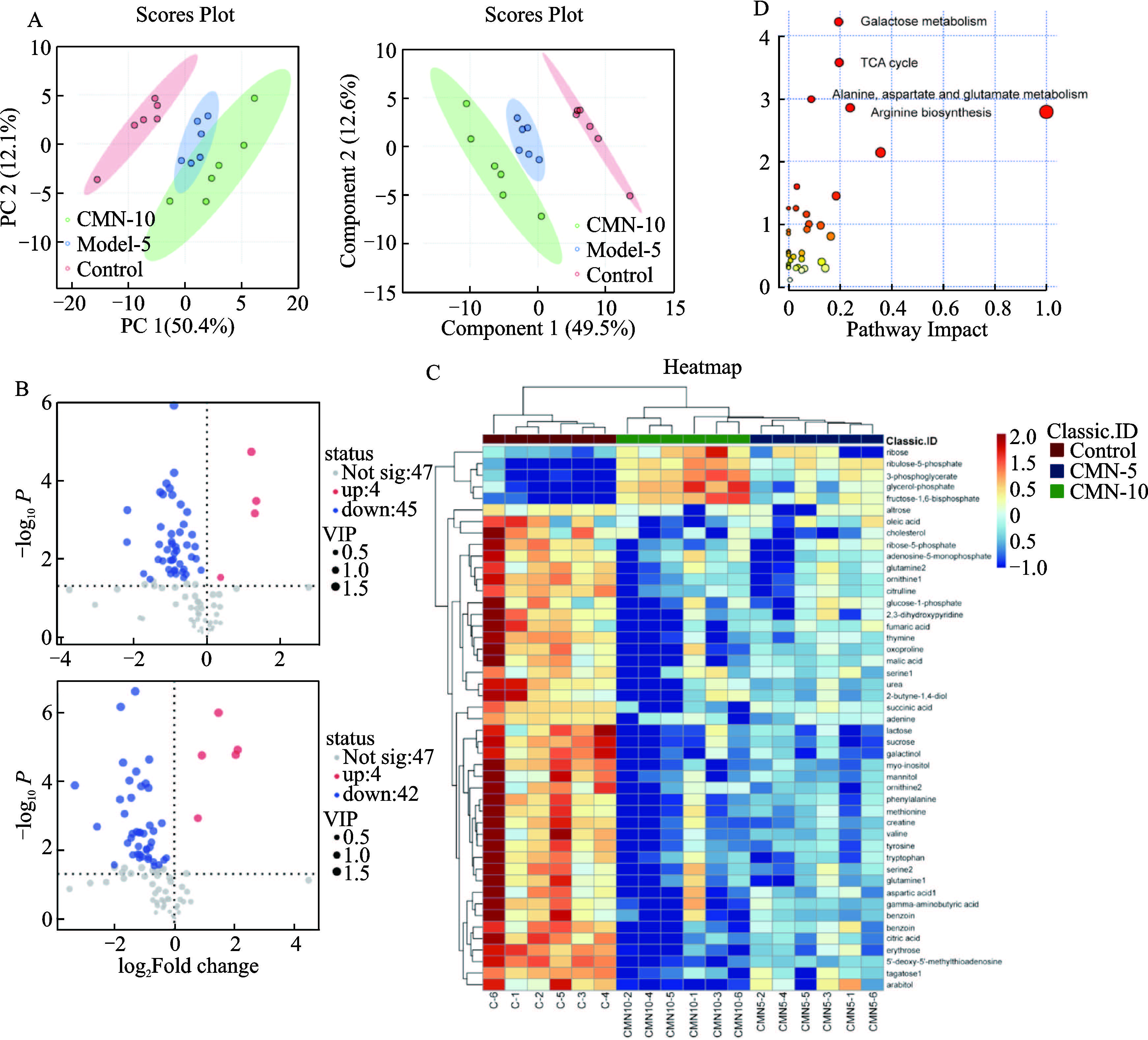
Note: A. PCA and PLS-DA score scatter plots of cell metabolites; B. Volcanic map of differential metabolites; C. Heatmap displaying differentially expressed metabolites in the Control, CMN-5, and CMN-10 groups; D. KEGG enrichment analysis for metabolic pathways in tumor samples
Fig. 6 The mechanism of CMN treating lung cancer was explored by cell metabolomics
4 Discussion
Lung cancer ranked among the primary causes of cancerassociated mortality globally, and its complex pathology greatly increases the difficulty of treatment[1]. Existing chemotherapeutic drugs for this malignancy exhibited multiple drawbacks,such as drug resistance, unsatisfied outcomes, and excessive expense. As a result, there was a need for new targeted therapeutic agents. Nowadays, TCM had gained recognition as an altherapeutic doses[9]. Nevertheless, the bioactive fractions and mechanisms of CM in managing lung cancer remained unknown. In this study, nucleotide and polysaccharide fractions were successfully extracted from CM. Animal experiments showed that CMN significantly reduced tumor volume and weight in a xenograft mouse model and exhibited fewer adverse effects compared with CM. Histopathological sections visually confirmed the improvement in tumor tissue following CMN treatment. In summary, this study found that CMN was the primary active fraction of CM responsible for its therapeutic effects in lung cancer.
The chemical constituents of CMN were analyzed using the UPLC-LTQ-Qrbitrap X technique in positive ion mode, leading to the successful identification of 13 compounds, including cordycepin, pentostatin, inosine, and adenine. As a cytotoxic nucleoside analog, cordycepin has been demonstrated to exert antitumor effects in previous studies[15-16]. It enhanced NK cell cytotoxicity, and this immunomodulatory activity enabled targeted elimination of cancer cells[17]. Previous studies showed that cordycepin inhibited the progression of drug-resistant lung cancer by activating the AMPK signaling pathway[18]. Pentostatin, a Food and Drug Administration-approved first-line anticancer drug, was first identified in CM in 2017[19], with subsequent investigations into advanced biological strategies[20]. Notably, pentostatin in CM prevented cordycepin degradation and prolonged its half-life[19,21]. Co-administration of pentostatin with cordycepin maintained cordycepin levels, indicating a stabilizing effect of pentostatin on cordycepin[22]. Inosine was found to enhance tumor immunogenicity to exert beneficial anticancer effects[23]. Inosine was an alternative carbon source for glucose-restricted CD8+ T cells and could improve the antitumor efficacy of T cell therapy or immune checkpoint inhibitors[24-25]. Adenine exhibited antiproliferative activity against tumor cells by inducing apoptosis and cell cycle S-phase arrest[26-27]. It was shown that adenine exerts anti-lung cancer effects by regulating AMPK-related signaling to block the oncogenic PI3K/Akt pathway mediated by adenine nucleotide translocase 2 (ANT2) in non-small cell lung cancer[28]. In addition, adenine further activated PI3K-Akt signaling by activating cAMP and antioxidant stress responses[29-30]. Collectively, the present study suggested that these components could contribute significantly to the antitumor activity of CMN.
Metabolic reprogramming, a process that fosters rapid cellular growth and proliferation by modulating energy metabolism,is regarded as a hallmark of cancer cells[31]. Tumor cells autonomously alter their metabolism through various pathways required for proliferation and survival [32]. Importantly, metabolomics aligns well with the holistic view of TCM. Therefore, the mechanism by which CMN treats lung cancer was investigated using metabolomics. Metabolomic analyses of serum and tumor samples indicated that CMN might exert therapeutic effects on lung cancer via the TCA cycle. The TCA cycle was a central hub in cellular metabolism, and many metabolic pathways, including glycolysis and fatty acid metabolism, were interconnected with the TCA cycle[33]. Specifically, the TCA cycle affected tumor proliferation and invasion by modulating energy metabolism, biosynthesis, and redox balance[34]. Studies found that elevated levels of TCA cycle intermediates were associated with reduced survival rates in lung cancer patients[35]. In lung cancer cells, inhibition of the TCA cycle led to lactate accumulation, which facilitated the formation of an immunosuppressive tumor microenvironment[36]. The TCA cycle tended to be dysregulated to satisfy the heightened energy requirements of cancer cells, and targeting the TCA cycle represented a possible therapeutic approach for treating cancer[37].
In vitro experiments further confirmed that CMN exerted a significant inhibitory effect on lung cancer cells, with this effect being concentration-dependent. CMN induced apoptosis in lung cancer cells, reduced their clonogenic capacity, inhibited wound closure rate, and arrested the cell cycle at the G1 phase,thereby suppressing cell proliferation and migration. These results suggested that CMN is a potential anti-lung cancer therapy. Metabolomic analyses of tumor cell samples further confirmed that CMN exerted its therapeutic effects on lung cancer via the TCA cycle. Previous studies showed that malic acid,a key intermediate in the TCA cycle, accumulated in lung cancer tumors[38]. Additionally, malate dehydrogenase, a biomarker for tumor prognosis, was upregulated in various cancers and contributed to the development of tumor resistance through multiple mechanisms[39]. In summary, the present study further validated that CMN inhibited lung cancer via the TCA cycle from the perspective of cellular metabolomics.
In this study, nucleotide and polysaccharide fractions were successfully extracted from CM. A total of 13 components were identified in CMN. The efficacies of fractions in CM in alleviating lung cancer were evaluated. The growth of tumors in xenograft nude mice and the proliferation of tumor cells were inhibited by CMN, with fewer side effects. The mechanism by which CMN treated lung cancer was investigated from multiple perspectives using metabolomics for the first time, both in vivo and in vitro. The current work found that CMN treated lung cancer by regulating the TCA cycle pathway. Therefore, this study might provide a theoretical basis for the application of CM in lung cancer treatment.
References:
[1] LEITER A, VELUSWAMY R R, WISNIVESKY J P. The global burden of lung cancer: Current status and future trends[J]. Nat Rev Clin Oncol, 2023, 20(9): 624-639.
[2] SHIELDS M D, LOVLY C M. Refining neoadjuvant immunotherapy for resectable lung cancer[J]. Nat Med, 2024, 30(6):1535-1536.
[3] LI Y T, YAN B S, HE S M. Advances and challenges in the treatment of lung cancer[J]. Biomed Pharmacother, 2023, 169:115891.
[4] XIANG L, GAO Y, CHEN S Y, et al. Therapeutic potential of Scutellaria baicalensis Georgi in lung cancer therapy[J]. Phytomedicine, 2022, 95: 153727.
[5] ZHANG W, ZONG Y Q, HUANG X, et al. Cordyceps militaris alleviates COPD by regulating amino acid metabolism, gut microbiota and short chain fatty acids[J]. J Ethnopharmacol, 2025,346: 119701.
[6] XU C H, WU F F, ZOU Z C, et al. Discovery of the chemical constituents, structural characteristics, and pharmacological functions of Chinese caterpillar fungus[J]. Open Chem, 2023, 21:20220337.
[7] KRISHNA K V, ULHAS R S, MALAVIYA A. Bioactive compounds from Cordyceps and their therapeutic potential[J]. Crit Rev Biotechnol, 2024, 44(5): 753-773.
[8] ZENG J P, ZHOU Y, LYU M D, et al. Cordyceps militaris: A novel mushroom platform for metabolic engineering[J]. Biotechnol Adv, 2024, 74: 108396.
[9] LU Y Y, HUANG X, LUO Z C, et al. Mechanism of Cordyceps militaris against non-small cell lung cancer: Based on serum metabolomics[J]. China J Chin Mater Med, 2022, 47(18): 5032-5039.
[10] QU J M, KE F, LIU Z R, et al. Uncovering the mechanisms of dandelion against triple-negative breast cancer using a combined network pharmacology, molecular pharmacology and metabolomics approach[J]. Phytomedicine, 2022, 99: 153986.
[11] SHI W B, WANG Z X, LIU H B, et al. Study on the mechanism of Fufang E’jiao Jiang on precancerous lesions of gastric cancer based on network pharmacology and metabolomics[J]. J Ethnopharmacol, 2023, 304: 116030.
[12] LONG F, PU X Y, WANG X, et al. A metabolic fingerprint of ovarian cancer: A novel diagnostic strategy employing plasma EVbased metabolomics and machine learning algorithms[J]. J Ovarian Res, 2025, 18(1): 26.
[13] PENG L J, ZHANG K, LI Y J, et al. Real-world evidence of traditional Chinese medicine (TCM) treatment on cancer: A literature-based review[J]. Evid Based Complement Alternat Med, 2022, 2022: 7770380.
[14] SONG S Y, PARK J H, PARK S J, et al. Synergistic effect of HAD-B1 and afatinib against gefitinib resistance of non-small cell lung cancer [J]. Integr Cancer Ther, 2022, 21:
15347354221144311.
[15] KHAN M A, TANIA M. Cordycepin and kinase inhibition in cancer[J]. Drug Discov Today, 2023, 28(3): 103481.
[16] SERPI M, FERRARI V, MCGUIGAN C, et al. Synthesis and characterization of NUC-7738, an aryloxy phosphoramidate of 3'-deoxyadenosine, as a potential anticancer agent[J]. J Med Chem, 2022, 65(23): 15789-15804.
[17] THEPMALEE C, JENKHAM P, RAMWARUNGKURA B, et al. Enhancing cancer immunotherapy using cordycepin and Cordyceps militaris extract to sensitize cancer cells and modulate immune responses[J]. Sci Rep, 2024, 14(1): 21907.
[18] WEI C L, YAO X J, JIANG Z B, et al. Cordycepin inhibits drugresistance non-small cell lung cancer progression by activating AMPK signaling pathway[J]. Pharmacol Res, 2019, 144:79-89.
[19] XIA Y L, LUO F F, SHANG Y F, et al. Fungal cordycepin biosynthesis is coupled with the production of the safeguard molecule pentostatin[J]. Cell Chem Biol, 2017, 24(12): 1479-1489. e4.
[20] ZOU G, LI B, WANG Y, et al. Efficient conversion of spent mushroom substrate into a high value-added anticancer drug pentostatin with engineered Cordyceps militaris[J]. Green Chem,2021, 23(24): 10030-10038.
[21] TURK A, LEE S, YEON S W, et al. Adenosine deaminase inhibitory activity of medicinal plants: Boost the production of cordycepin in Cordyceps militaris[J]. Antioxidants (Basel),2023, 12(6): 1260.
[22] LEE J B, RADHI M, CIPOLLA E, et al. A novel nucleoside rescue metabolic pathway may be responsible for therapeutic effect of orally administered cordycepin[J]. Sci Rep, 2019, 9(1):15760.
[23] ZHANG L, JIANG L, YU L, et al. Inhibition of UBA6 by inosine augments tumour immunogenicity and responses[J]. Nat Commun, 2022, 13: 5413.
[24] WANG T T, RASHIDA GNANAPRAKASAM J N, CHEN X Y,et al. Inosine is an alternative carbon source for CD8+-T-cell function under glucose restriction[J]. Nat Metab, 2020, 2(7):635-647.
[25] HU Y, SARKAR A, SONG K, et al. Selective refueling of CART cells using ADA1 and CD26 boosts antitumor immunity[J]. Cell Rep Med, 2024, 5(5): 101530.
[26] YUAN Z N, LI Y P, ZHANG S F, et al. Extracellular matrix remodeling in tumor progression and immune escape: From mechanisms to treatments[J]. Mol Cancer, 2023, 22(1): 48.
[27] SU W W, HUANG J Y, CHEN H M, et al. Adenine inhibits growth of hepatocellular carcinoma cells via AMPK-mediated S phase arrest and apoptotic cascade[J]. Int J Med Sci, 2020, 17(5): 678-684.
[28] JANG JY, KIM YG, NAM SJ, et al. Targeting adenine nucleotide translocase-2 (ANT2) to overcome resistance to epidermal growth factor receptor tyrosine kinase inhibitor in non-small cell lung cancer[J]. Mol Ca Therap, 2016, 15(6): 1387-1396.
[29] HAO J F, ZHANG X Y, HU R X, et al. Metabolomics combined with network pharmacology reveals a role for astragaloside IV in inhibiting enterovirus 71 replication via PI3K-AKT signaling[J]. J Transl Med, 2024, 22(1): 555.
[30] FENG S J, TANG D, WANG Y C, et al. The mechanism of ferroptosis and its related diseases[J]. Mol Biomed, 2023, 4(1): 33.
[31] LIU S, ZHANG X D, WANG W Z, et al. Metabolic reprogramming and therapeutic resistance in primary and metastatic breast cancer[J]. Mol Cancer, 2024, 23(1): 261.
[32] MARTÍNEZ-REYES I, CHANDEL N S. Cancer metabolism:Looking forward[J]. Nat Rev Cancer, 2021, 21(10): 669-680.
[33] CHELAKKOT C, CHELAKKOT V S, SHIN Y, et al. Modulating glycolysis to improve cancer therapy[J]. Int J Mol Sci, 2023, 24(3): 2606.
[34] BARTMAN C R, WEILANDT D R, SHEN Y H, et al. Slow TCA flux and ATP production in primary solid tumours but not metastases[J]. Nature, 2023, 614(7947): 349-357.
[35] CAI L, HAMMOND N G, TASDOGAN A, et al. High glucose contribution to the TCA cycle is a feature of aggressive non–small cell lung cancer in patients[J]. Cancer Discov, 2025, 15(4):702-716.
[36] QIAN Y, GALAN-COBO A, GUIJARRO I, et al. MCT4-dependent lactate secretion suppresses antitumor immunity in LKB1-deficient lung adenocarcinoma[J]. Cancer Cell, 2023, 41(7):1363-1380.
[37] YANG J Q, CHEN F H, FU Z Z, et al. Blocking the TCA cycle in cancer cells potentiates CD36+ T-cell-mediated antitumor immunity by suppressing ER stress-associated THBS2 signaling[J]. Cancer Res, 2025, 85(11): 2014-2026.
[38] SAINERO-ALCOLADO L, LIANO-PONS J, RUIZ-PEREZ M V, et al. Targeting mitochondrial metabolism for precision medicine in cancer[J]. Cell Death Differ, 2022, 29(7): 1304-1317.
[39] WANG H, CUI W L, YUE S, et al. Malic enzymes in cancer:Regulatory mechanisms, functions, and therapeutic implications[J]. Redox Biol, 2024, 75: 103273.
(编辑:董宇)