基于非靶向代谢组学技术研究蛹虫草核苷的抗肺癌机制
柴晓倩1,2,陆颖颖1,2,陈雨雨1,2,罗子宸1,2,3,单进军3,张雯1,2,狄留庆1,2
(1. 南京中医药大学药学院,江苏 南京 210023;2. 江苏省中药高效给药系统工程技术研究中心,江苏 南京 210023;3. 南京中医药大学医学代谢组学中心,江苏 南京 210023)
摘要:
目的 筛选蛹虫草发挥抗肺癌作用的有效组分,采用非靶向代谢组学技术研究蛹虫草核苷对肺癌荷瘤裸鼠及肺癌细胞内源性代谢物的影响,并探讨其潜在作用机制。
方法 提取并纯化蛹虫草核苷和多糖组分,采用UPLC-LTQ-Orbitrap X技术对蛹虫草核苷进行化学成分分析。构建人肺癌裸鼠移植瘤模型,分别用蛹虫草及不同组分提取物干预2周,通过肿瘤的体积与质量、组织病理学观察筛选蛹虫草抗肺癌作用的活性组分。收集裸鼠血清及肿瘤组织样本,利用GC-MS技术进行非靶向代谢组学分析。通过CCK-8、集落形成、划痕愈合试验及细胞周期分析,评价蛹虫草核苷组分对肺癌细胞的影响。通过细胞代谢组学分析验证蛹虫草核苷发挥抗肺癌作用的相关代谢通路。
结果 从蛹虫草核苷中鉴定出虫草素、肌苷、腺嘌呤等13种化学成分。蛹虫草核苷能显著抑制人肺癌荷瘤裸鼠肿瘤生长,且毒副作用较小;在细胞水平上能抑制肺癌细胞的增殖、迁移与周期进程。血清代谢组学分析共鉴定出38种差异代谢物,肿瘤组织代谢组学分析共鉴定出44种差异代谢物,上述差异代谢物主要参与三羧酸循环通路。肿瘤细胞代谢组学分析鉴定的差异代谢物进一步证实了该结论。
结论 蛹虫草核苷是蛹虫草发挥抗肺癌作用的有效组分,可通过调控三羧酸循环通路发挥抗肺癌作用。
关键词: 蛹虫草;肺癌;三羧酸循环;非靶向代谢组学;核苷
中图分类号:R285. 5
文献标志码:A
文章编号:1672-0482(2026)06-0831-11
DOI:10. 14148/j. issn. 1672-0482. 2026. 0831
引文格式:柴晓倩,陆颖颖,陈雨雨,等. 基于非靶向代谢组学技术研究蛹虫草核苷的抗肺癌机制[J]. 南京中医药大学学报,2026,42(6):831-841.
Investigation on the Anti-Lung Cancer Mechanism of Cordyceps Militaris Nucleosides by Non-Targeted Metabolomics
CHAI Xiaoqian1,2, LU Yingying1,2, CHEN Yuyu1,2, LUO Zichen1,2,3, SHAN Jinjun3, ZHANG Wen1,2, DI Liuqing1,2
(1. School of Pharmacy, Nanjing University of Chinese Medicine, Nanjing 210023, China; 2. Jiangsu Engineering Research Center for Efficient Delivery System of TCM, Nanjing 210023, China; 3. Medical Metabolomics Center, Nanjing University of Chinese Medicine, Nanjing 210023, China)
ABSTRACT:
OBJECTIVE To screen the active fraction of Cordyceps militaris (CM) exerting anti-lung cancer effects, investigate its effects on endogenous metabolites in lung cancer-bearing nude mice and explore the potential mechanism of action by non-targeted metabolomics.
METHODS The nucleosides (CMN) and polysaccharides (CMP) of CM were extracted and purified. The components in CMN were identified by UPLC-LTQ-Orbitrap X instrument. The model of human lung cancer xenograft in nude mice was established, and treated with CM and its different fraction extracts for two weeks. The active fraction of CM with anti-lung cancer effect was screened by monitoring tumor volume, tumor weight and histopathological alterations. Serum and tumor tissue samples were collected, and GC-MS technology was used for metabolomics analysis. The effects of CMN on lung cancer cells were evaluated by CCK-8, colony formation, wound healing assays and cell cycle analysis. The metabolic pathways involved in the anti-lung cancer effect of CMN were further verified by cellular metabolomics.
RESULTS A total of 13 components including cordycepin, inosine and adenine were identified from CMN. CMN significantly inhibited tumor growth with low toxicity. At the cellular level, CMN inhibited the proliferation, migration and cell cycle progression of lung cancer cells. A total of 38 differential metabolites were identified by serum metabolomics analyses, and 44 by tumor tissue metabolomics analyses, which were mainly involved in the citrate cycle (TCA cycle). This conclusion was further confirmed by differential metabolites identified through tumor cell metabolomics analyses.
CONCLUSION CMN is theactive fraction of CM exerting anti-lung cancer effects, which might exert anti-lung cancer effects by regulating the TCA cycle pathway.
KEYWORDS: Cordyceps militaris; lung cancer; TCA cycle; non-targeted metabolomics; nucleosides
收稿日期: 2025-11-22
基金项目: 江苏省高等学校自然科学研究项目(19KJB360004);江苏省研究生科研与实践创新计划项目(KYCX24_2297)
第一作者: 柴晓倩,女,硕士研究生,E-mail:c19106413709@163. com
通信作者: 张雯,女,副教授,主要从事中药药效及其物质基础研究,E-mail:wenzhang@njucm. edu. cn;
狄留庆,男,教授,主要从事中药高效给药系统设计与评价研究,E-mail:diliuqing@njucm. edu. cn
1 Introduction
Worldwide, lung cancer ranks as the top cause of cancerassociated deaths, with an exceptionally high fatality rate[1].While lung cancer can be treated with chemotherapy, surgery,radiotherapy, and immunotherapy, chemotherapy still stands as the most significant and commonly employed therapeutic approach for patients with this disease[2]. Third-generation chemotherapy drugs such as docetaxel and pemetrexed are usually chosen as postoperative adjuvant therapy for lung cancer. Yet,nearly all traditional medications have the same drawbacks,such as a lack of specific targeting, poor bioavailability, and the emergence of drug resistance[3]. As a result, it is urgent and essential to strive for alternative treatments for lung cancer with minimal side effects.
Compared with conventional therapies, Traditional Chinese Medicine (TCM) has the advantage of fewer side effects while achieving a similar effect, and improving patients' quality of life[4]. Cordyceps militaris (CM) is a representative species of Cordyceps mushrooms, which has been used as a lung and kidney tonic for treating chronic bronchitis, asthma, tuberculosis,and other respiratory diseases for thousands of years[5]. Compared with monomeric components, the active fractions are better at reflecting the holistic action characteristics of TCM. The fractions of CM are nucleosides (CMN), polysaccharides(CMP), sterols (CME), and various trace elements[6]. Through the intervention on the model of human lung cancer xenograft in nude mice, we systematically studied the anti-lung cancer effects of CM and its fractions, and screened CMN as the key research fraction. Notably, nucleosides has been the focus of CM in anti-tumor research, including cordycepin, pentostatin, uri⁃dine, adenosine, and hypoxanthine[7-8]. Our previous study demonstrated that CM extract significantly inhibited lung cancer[9].However, the active fractions of CM for the treatment of lung cancer and their related molecular mechanisms remain unclear.
Metabolomics enables the acquisition of extensive information on intracellular metabolite changes. The merits of metabolomics correspond to the holistic and systemic outlook of TCM theory, and in recent years, this analytical approach has grown into a widely adopted research method within TCM[10-11]. Serum is considered a suitable metabolic sample reflecting the overall metabolic status of the organism because all organs and tissues are filled with blood[12]. The tumor tissue and cell samples derived from lung cancer tissues and cell lines respectively could reflect the direct effect of the active ingredients of CM on lung cancer.
In this study, CMN and CMP were extracted and purified from CM firstly. The chemical compositions of CMN were analyzed by UPLC-LTQ-Orbitrap X. Afterwards, the anti-lung cancer effects of CM, CMN, CMP and sterol of CM (CME)were evaluated by the xenograft mice model. The molecular mechanism of CMN against lung cancer was investigated by serum and tumor tissues metabolomics. Moreover, the lung cancer-inhibiting properties of CMN were further assessed using CCK-8, colony formation, wound healing assays, and cell cycle analysis. Finally, the molecular mechanism of CMN against lung cancer was further validated by metabolomics of tumor cells.
2 Materials and methods
2. 1 Reagents
Cordyceps militaris was purchased from Hunan Yandi Bioengineering Co. , Ltd. (Hunan, China), and it was authenticated by Prof. Chen Jianwei from Nanjing University of Chinese Medicine. Macroporous resins including NKA-2 and D101 were purchased from Beijing Solarbio Science & Technology Co. ,Ltd. (Beijing, China). Uridine, cordycepin, adenosine,ergosterol, guanosine, inosine, cytidine, xanthine, adenine, 2'-deoxyguanosine, hypoxanthine and glucose controls were purchased from Shanghai Yuanye Bio-Technology Co. , Ltd.(Shanghai, China). Thymidine and 2'-deoxyadenosine were provided by National Institutes for Food and Drug Contro (Beijing, China). N6-(2-hydroxyethyl) adenosine was purchased from Sichuan Vicki Biotechnology Co. , Ltd. (Sichuan,China). Cis-diamminedichloroplatinum (DDP) was bought from Shanghai Aladdin Biochemical Technology Co. , Ltd.(Shanghai, China). F12K medium was purchased from Shanghai Basalmedia Technologies Co. , Ltd. (Shanghai, China).Fetal bovine serum (FBS) was purchased from Shanghai Nova Pharmaceutical Technology Co. , Ltd. (Shanghai, China).Penicillin and Trypsin-EDTA Solution were provided by Nanjing BioChannel Biotechnology Co. , Ltd. (Jiangsu, China).Cell Cycle Staining Kit was purchased from Hangzhou Lianke Biotechnology Co. , Ltd.( Zhejiang, China).
2. 2 Extraction and purification of CMN and CMP
CM powder was soaked in 20-fold water for 30 min and extracted via ultrasonic treatment for 60 min. After filtration, the water-soluble fraction was concentrated to a specific volume under reduced pressure. The resulting mixture was allowed to stand overnight at 4 ℃ and then washed three times with anhydrous ethanol. The alcohol-soluble components were fixed with 80% ethanol to obtain the CMN crude extract. The alcoholinsoluble ingredients were fixed with pure water to obtain the CMP crude extract. Next, a 190 mL CMN solution( 15 mg·mL-1)was loaded onto an NKA-2 resin column and eluted with 60% ethanol. In addition, a 100 mL CMP solution (10 mg·mL-1)was purified using an X-5 resin column. The content of CMN and CMP was determined by HPLC and UV, respectively.
2. 3 Components identification of CMN by UPLC-LTQOrbitrap X
Preparation of control solution: appropriate amounts of cordycepin, xanthine, uridine, inosine, adenine, thymidine,adenosine, N6-(2-hydroxyethyl) adenosine, guanosine, cytidine, 2'-deoxyadenosine, and 2'-deoxyguanosine were each precisely weighed and dissolved in 70% methanol to prepare stock standard solutions. Subsequently, the solutions were centrifuged at 18 000 r·min-1 for 10 minutes. The supernatant was then collected and filtered through a 0. 22 μm membrane. Preparation of test solution: 5 mg of CMN freeze-dried powder was dissolved in 70% methanol, centrifuged, and analyzed using UPLC-LTQ-Orbitrap X. The instrument conditions were described in detail in the supplementary materials.
2. 4 Animal experiments
The experiment was approved by the Ethics Committee of Animal Experimentation of Nanjing University of Chinese Medicine on July 20, 2022, with ethical application No. 202207A008. Male Balb/c nude mice (6 weeks) were purchased from Shanghai SLAC Laboratory Animal Co. , Ltd.(Shanghai, China). To establish a subcutaneous xenograft model, 48 nude mice were subcutaneously injected with 0. 2 mL of A549 cell suspension (3×10⁷ mL-1), while 8 nude mice were injected with 0. 2 mL of PBS as a blank control group. Once the tumor volume reached approximately 50 mm³, the xenograft mice were randomly divided into six groups. Mice in the blank and model groups were orally administered saline daily. Mice in the CM, CMN, CMP, and CME groups were orally administered the corresponding drug (1 g crude herb·kg-1) for 14 consecutive days. Mice in the DDP group were intraperitoneally injected with DDP (4 mg·kg-1) every other day. After 2 weeks of treatment, blood was collected from the orbits of the xenograft mice for metabolomics analysis. Lung, heart, spleen, liver, kidney,and tumor tissues were collected for pathological analysis.
2. 5 Cell phenotyping assays
2. 5. 1 CCK-8 assay
A549 cells were plated in 96-well plates at a density of 5 000 cells·well-1. Subsequently, the medium was substituted with fresh medium containing varying concentrations of CMN. After 24, 48, and 72 hours of incubation,10% CCK-8 reagent was added to each well, and the plates were further incubated at 37° C for 1 h. The absorbance was measured at 450 nm using a microplate reader (PerkinElmer,USA). Cell viability and the half-maximal inhibitory concentration( IC50) were then calculated.
2. 5. 2 Colony formation assay
A549 cells were inoculated into 6-well plates at a density of 500 cells per well. Subsequently, the cells were incubated for 24 hours in medium supplemented with varying concentrations of CMN (0, 0. 5, 1,2 μg·mL-1). During colony development, the medium was refreshed every 3 days, and cell growth was monitored daily via microscopy. Following the incubation period, the cells were rinsed, fixed with formaldehyde solution, stained, and airdried.Colonies were visualized and photographed under a microscope. Colonies containing ≥50 cells were counted, and the relative proliferative survival rate and colony formation inhibition rate were calculated.
2. 5. 3 Wound healing assay
A549 cells were seeded into 6-well plates at a density of 5×10 ⁵ cells·well-1. Once the cells achieved 100% confluence, a sterile 200 μL pipette tip was used to create a vertical scratch across the cell monolayer. Postscratching,the cells were incubated in medium containing various concentrations of CMN (0, 2, 4, 8 μg·mL-1). The scratch closure was monitored under an inverted microscope, and images were captured at designated time points.
2. 5. 4 Cell cycle assay
A549 cells were plated into 6-well plates at a density of 5×10⁵ cells·well-1. After overnight incubation to allow cell attachment, the medium was aspirated and replaced with fresh medium containing varying concentrations of CMN (0, 0. 5, 1, 2 μg·mL-1). Following a 24-hour treatment period, the cells were harvested, permeabilized with 5 μL of permeabilization solution, and stained with 500 μL of DNA staining solution for 1 hour at room temperature. The stained cells were filtered through a 300-mesh nylon strainer to remove cell clumps and analyzed by flow cytometry. Data acquisition and analysis were performed using Kaluza Analysis 2. 1 software.
2. 6 Metabolomic analysis
Precisely pipet 50 μL of each serum sample, followed by the addition of 200 μL methanol solution containing 1,2-¹³C myristic acid (12. 5 μg·mL-1). The mixture was vortexed and then centrifuged at 4 °C and 18 000 r·min-1 for 10 minutes. The supernatant was collected and transferred to a centrifugal concentrator for evaporation to dryness. Subsequently, 30 μL of a pyridine solution with 10 mg·mL-1 oxalyl chloride was added. The mixture was vortexed and then oscillated at 300 r·min-1 for 90 min at 30 °C. After that, 30 μL of BSTFA was added, and the mixture was vortexed before being shaken at 37 °C and 300 r·min-1 for 30 minutes. It was then centrifuged at 18 000 r·min-1 for 10 minutes, and finally, GC-MS analysis was performed.
Tumor tissue samples were placed on ice, and 20 mg of each tissue sample was accurately weighed. Then, 500 μL of methanol was added for homogenization. A 350 μL aliquot of the tumor tissue homogenate was extracted, and 10 μL of anice-cold methanol solution containing 1,2- ¹³C myristic acid(300 μg·mL-1) was added. The mixture was vortexed for 3 min and centrifuged at 4 ℃ and 18 000 r·min-1 for 10 min. A 150 μL portion of the supernatant was evaporated to dryness using a centrifugal concentrator. Afterward, 50 μL of a pyridine solution with 10 mg·mL-1 methoxylamine hydrochloride was added, followed by 50 μL of BSTFA. The mixture was vortexed for 5 minutes and then incubated at 37 ℃ and 300 r·min-1 for 30 min. After centrifugation at 18 000 r·min-1 for 10 min, performed GC-MS analysis.
A549 cells were seeded in 6-well plates at a density of 1×10⁶ cells per well. After the cells had adhered, medium containing drugs at different concentrations (0, 5, 10 μg·mL-1 CMN) was added. Following 24 h of incubation, the cells were resuspended in 500 μL of ice-cold methanol solution containing 1,2-¹³C myristic acid( 5 μg·mL-1) and centrifuged at 18 000 r·min-1 for 10 min. A 400 μL aliquot of the supernatant was evaporated to dryness in a centrifugal concentrator. Then, 30 μL of pyridine containing 10 mg·mL-1 methoxylamine hydrochloride was added, and the mixture was vortexed for 5 min and oscillated at 30 ℃ and 300 r·min-1 for 90 min. Finally, 30 μL of BSTFA was added, and the mixture was vortexed for 5 min before being shaken at 30 ℃ and 300 r·min-1 for 30 min. After centrifugation at 18 000 r·min-1 for 10 min, GC-MS analysis was performed.
Detailed descriptions of the GC-MS conditions were provided in the supplementary materials.
3 Results
3. 1 Components identification of CMN
The chemical components of CMN were analyzed using UPLC-LTQ-Q Orbitrap X in positive ion mode. The total ion chromatograms (TICs) were shown in Fig. 1A and Fig. 1B. The structural elucidation of chemical components was determined using reference compounds, chromatographic retention times,and MS/MS datasets. A total of 13 compounds were identified inCMN, including adenosine, inosine, 2'-deoxyguanosine,2'-deoxyadenosine, L-guanosine, N6-(2-hydroxyethyl) adenosine, cordycepin, pentostatin, cytidine, uridine, thymidine, adenine, and xanthine. The precise mass readings for the protonated molecular ions of the 13 CMN components under positive ion mode were summarized in Table S1, encompassing retention times, molecular formulas, experimental and theoretical masses, mDa and ppm errors.
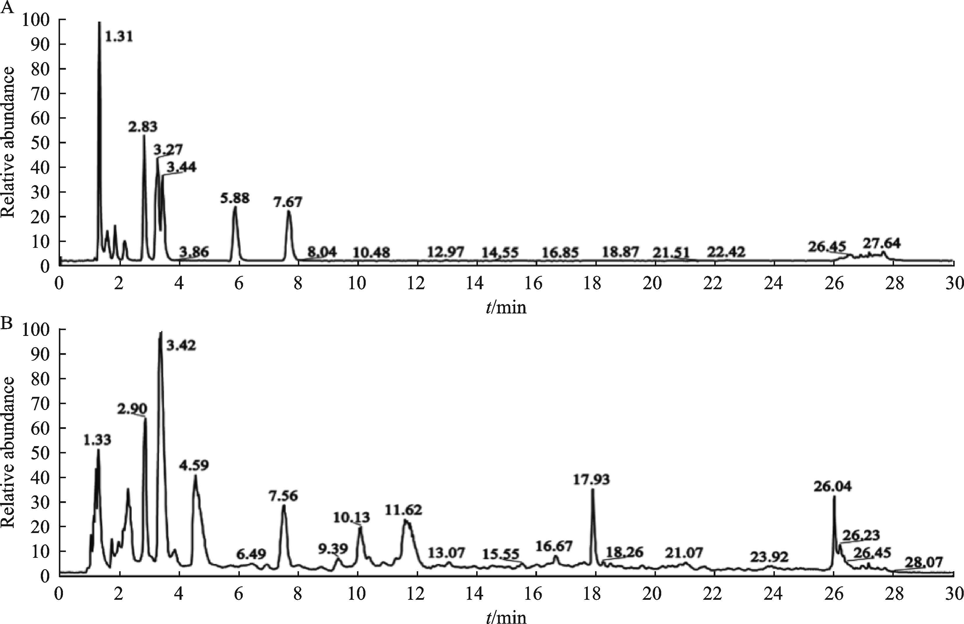
Note: A. Total ion chromatography of the mixed control substances; B. Total ion chromatography of CMN
Fig. 1 The constituents of CMN identified by UPLC-LTQ-Orbitrap X