接第一篇:
Deacetylase activity assays, performed using a fluorescent AMC substrate, revealed that all ΔMrsir2 mutant strains exhibited significantly reduced deacetylase activity compared to the wild-type strain (WT) (Fig 2A). Notably, the deacetylase activity of the ΔMrsir2–3 strain was lower than that of the other three mutant strains, indicating that MrSIR2–3 plays a crucial role in fungal deacetylation processes.
Phenotypic assays conducted on Potato Dextrose Agar (PDA) demonstrated that deletion of Mrsir2 genes did not impact fungal growth diameters, suggesting that Mrsir2 genes do not regulate overall mycelial growth under these conditions (Fig 2B). Assessments of conidial yields revealed substantial reductions in conidial production across all ΔMrsir2 mutants compared to the WT strain. Specifically, Deletion of Mrsir2–1, Mrsir2–2, Mrsir2–3, and Mrsir2–4 exhibited reductions in conidial production by 97%, 66%, 92%, and 84%, respectively (Fig 2C). Consistent results were obtained from conidiation analyses performed on different media and at various time points, with Mrsir2 mutants consistently showing significantly lower conidial production than the WT strain (S3 Fig). Complementation of these mutants restored conidiation to levels comparable to the WT strain, thereby confirming the essential roles of Mrsir2 genes in conidiation. Transcriptomic analysis of conidiation-associated genes further indicated that deletion of Mrsir2 genes leads to significant downregulation of key regulatory genes, such as stc, hymA, flbC, phiA, fadA, flbD, sakA, abaA, brlA, stuA, medA, and wetA, during conidiation (S4 Fig).
Collectively, these findings demonstrate that the MrSIR2 proteins exhibit functional deacetylase activity and play essential roles in efficient conidial production in M. robertsii.
Deacetylase MrSIR2–3 deacetylates MrATG4
Given the elevated expression of Mrsir2–3 during conidiation and its cytoplasmic localization, combined with its major role in deacetylation at the assayed stage and significant impact on conidial production, we prioritized Mrsir2–3 for further investigation. Using yeast two-hybrid screening with pGBKT7-MrSIR2–3 as bait, we identified 24 potential interacting proteins based a genome-wide screening, including MrATG4, a key autophagy-related protein with a molecular weight of 28.4 kDa (S1 Table).
Further yeast two-hybrid assays revealed that MrSIR2–3 specifically interacts with MrATG4 but not with other components of the ATG8-conjugation system (Fig 3A). To validate this interaction, we conducted bimolecular fluorescence complementation (BiFC) and co-immunoprecipitation (co-IP) assays. Both methods confirmed the physical association between MrSIR2–3 and MrATG4 (Figs 3B, 3C, S5 and S6).
To assess the levels of MrATG4 acetylation, we constructed and harvested MrATG4-FLAG strains of the WT and ΔMrsir2–3 mutant at early conidiation (2.5 days). FLAG immunoprecipitates were immunoblotted with an anti-pan-acetylated lysine antibody, revealing higher MrATG4 acetylation levels in the WT strain compared to the ΔMrsir2–3 mutant (Fig 3D). These findings indicate that MrSIR2–3 directly interacts with and deacetylates MrATG4, thereby modulating its acetylation state during conidiation.
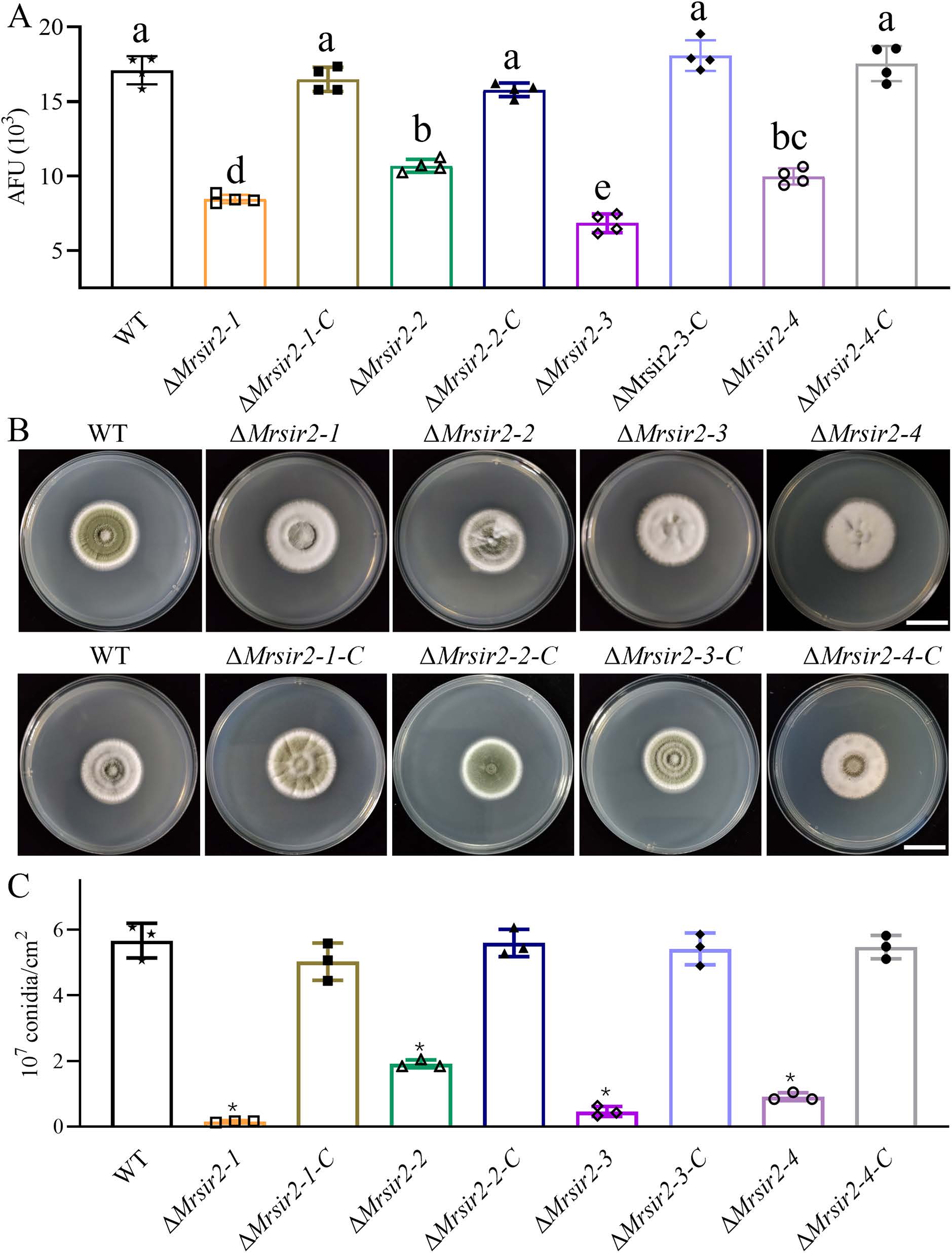
Fig 2. Contribution of MrSIR2s to conidial yields and deacetylase activity. (A) Intracellular deacetylase activity in different strains measured in arbitrary fluorescence units (AFU). * p < 0.01. (B) Colony phenotypes of WT and mutant strains on PDA media after 7 days of growth at 25 °C. (C) Quantification of conidial yields from different strains after two weeks of growth on PDA. * p < 0.01.
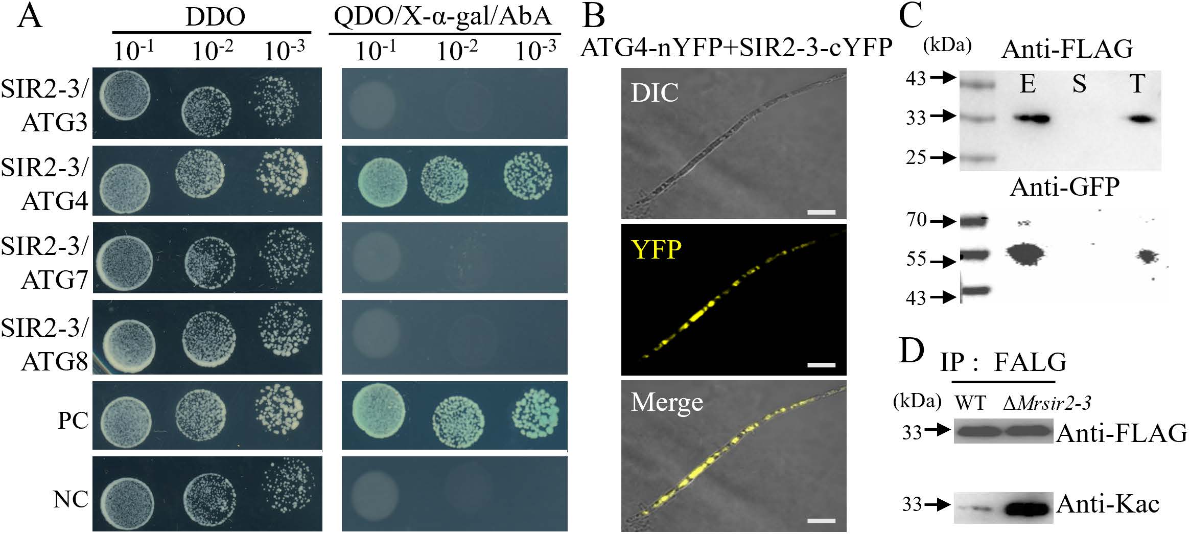
Fig 3. Interaction and deacetylation between MrSIR2-3 and MrATG4 in M. robertsii. (A) Yeast two-hybrid analysis demonstrating the interaction between MrSIR2-3 and the ATG8-conjugation system proteins. DDO, SD-T-L; QDO, SD-T-L-A-H. (B) Bimolecular fluorescence complementation (BiFC) assays for detecting in vivo protein interactions. Scale bar: 5 μm. (C) Co-immunoprecipitation assays. Total proteins, suspensions, and proteins eluted from anti-FLAG agarose from transformants co-expressing MrSIR2-3-GFP and MrATG4-FLAG. The blots were probed with anti-FLAG or anti-GFP antibodies. T, total; S, suspensions; E, elution. (D) FLAG co-IP and anti-acetyl-lysine immunoblot analysis of WT vs. ΔMrsir2-3 strains grown on PDA (2.5 d).
Regulation of autophagy by MrSIR2–3 and MrATG4 during conidiation
To elucidate the roles of MrSIR2–3 and MrATG4 in autophagy regulation, we utilized a GFP-MrATG8 autophagy marker and introduced it into WT, ΔMrsir2–3, and ΔMratg4 mutant strains. In the WT, autophagosomes and GFP-MrATG8 fluorescence were readily observed during the initial stages of conidiation (Fig 4A and 4B). In contrast, the ΔMrsir2–3 mutant exhibited an increased accumulation of autophagosomes and heightened GFP-MrATG8 fluorescence within the cytoplasm, indicative of elevated autophagic activity (Figs 4A–4D, S7 and S8). Conversely, the ΔMratg4 strain displayed minimal autophagosomes and faint GFP-MrATG8 fluorescence, suggesting impaired autophagy (Fig 4E and 4F). Transmission electron microscopy (TEM) further corroborated these observations, showing an abundance of autophagic bodies within vacuoles of the ΔMrsir2–3 mutant and a marked absence of autophagic bodies in ΔMratg4 (Fig 4G).
These results demonstrate that MrSIR2–3 acts as a negative regulator of autophagy, likely by deacetylating MrATG4, which is essential for proper autophagic function during conidiation.
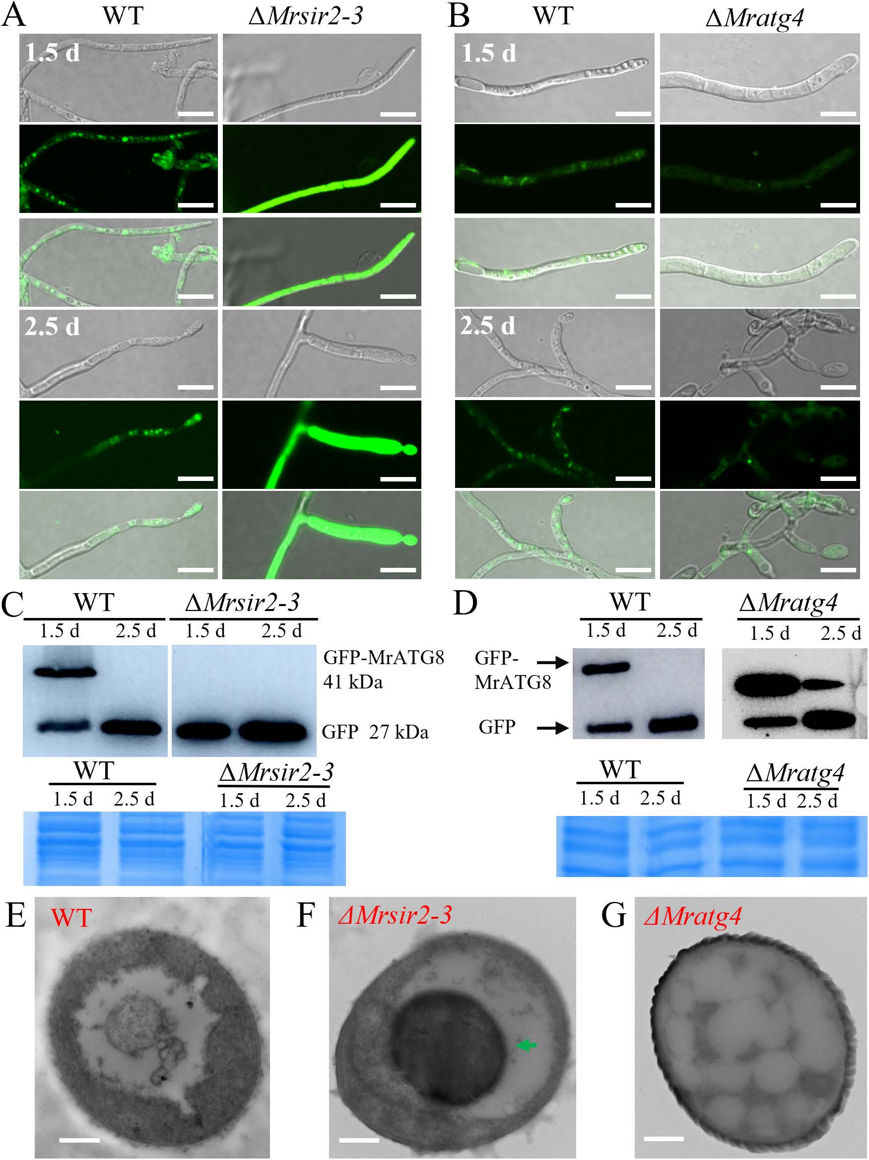
Fig 4. Critical roles of Mrsir2-3 and Mratg4 in autophagy of M. robertsii. (A and B) Cellular localization of autophagosomes during the initial mycelial growth (1.5 d) and initial conidia production stages (2.5 d). Scale bar: 10 μm. (C and D) Analysis of GFP-MrATG8 degradation in the WT and mutant mycelial cells. Immunoblotting was performed with anti-GFP. For each analysis, parallel protein gels were stained as references. (E - G) Transmission Electron Microscopy (TEM) analysis of the vacuolar localization features of Autophagosomes in the mycelial structures during conidiation in both wild-type WT and mutant strains. Autophagic bodies are indicated by arrows in the ΔMrsir2-3 cell. Bar: 2 μm.
Acetyltransferase MrKAT1 interacts with MrATG4
To identify potential acetyltransferases that interact with MrATG4, we performed yeast two-hybrid assays using a panel of acetyltransferase homologs from S. cerevisiae (Gcn5 (MAA_05172), Esa1 (MAA_02727), Hat1 (MAA_05971), Rtt109 (MAA_01374), Sas2 (MAA_04679), and Sas3 (MAA_02282) as bait. This screen identified the Hat1 homolog MAA_05971 as a specific interacting partner of MrATG4 (Fig 5A). We therefore designated this protein MrKAT1. To validate this interaction, we conducted co-IP assays. Co-expression of MrATG4-FLAG and MrKAT1-GFP in the wild-type (WT) strain resulted in the co-precipitation of MrKAT1 with MrATG4 using anti-FLAG beads, confirming their physical association (Fig 5B). Additionally, bimolecular fluorescence complementation (BiFC) assays were performed to confirm the in vivo interaction between MrKAT1 and MrATG4. The presence of YFP fluorescence indicated that MrKAT1 and MrATG4 interact in living cells (Figs 5C, S5 and S6).
Role of Mrkat1 in autophagy and conidiation
Mrkat1 deletion mutant and a complemented strain (ΔMrkat1-C) were generated to investigate the role of MrKAT1 in autophagy and conidiation (Figs 6 and S9). Phenotypic analysis revealed that the ΔMrkat1 mutant exhibited a significant reduction in conidial production compared to WT and ΔMrkat1-C (Fig 6A and B). Autophagic flux assessments using GFP-MrATG8 indicated that the ΔMrkat1 mutant displayed reduced autophagy levels, similar to the ΔMratg4 mutant (Fig C–E and S9). Immunoblotting further quantified autophagic flux by assessing the ratio of free GFP to total GFP-MrATG8, revealing a marked decrease in free GFP in ΔMrkat1 (Fig C–E). TEM analysis confirmed the reduced presence of autophagic bodies in ΔMrkat1, akin to ΔMratg4 (Fig 6F).
These findings establish MrKAT1 as a positive regulator of autophagy, functioning antagonistically to MrSIR2–3, and essential for efficient conidiation in M. robertsii.
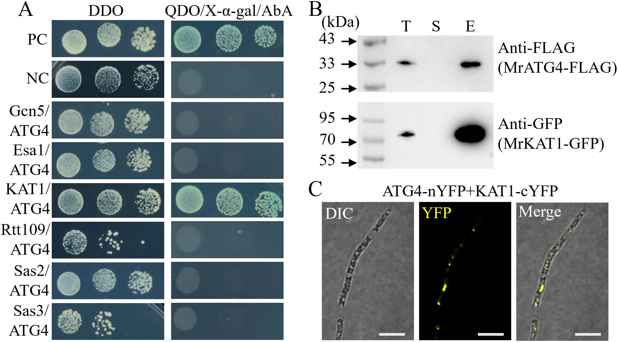
Fig 5. MrKAT1 interacted with MrATG4. (A) Yeast two-hybrid analysis of the interaction between MrATG4 and the Nε-lysine acetyltransferase proteins. DDO, SD-T-L; QDO, SD-T-L-A-H. (B) Co-IP assays. Western blots of total proteins, suspensions and proteins eluted from anti-FLAG agarose from transformants co-transformed MrKAT1-GFP and MrATG4-FLAG, MrATG4-FLAG and MrKAT1-GFP were detected with anti-FLAG or anti-GFP antibodies. T, total; S, suspensions; E, elution. (C) BiFC assays for detecting in vivo protein interactions. Scale bar: 5 μm.
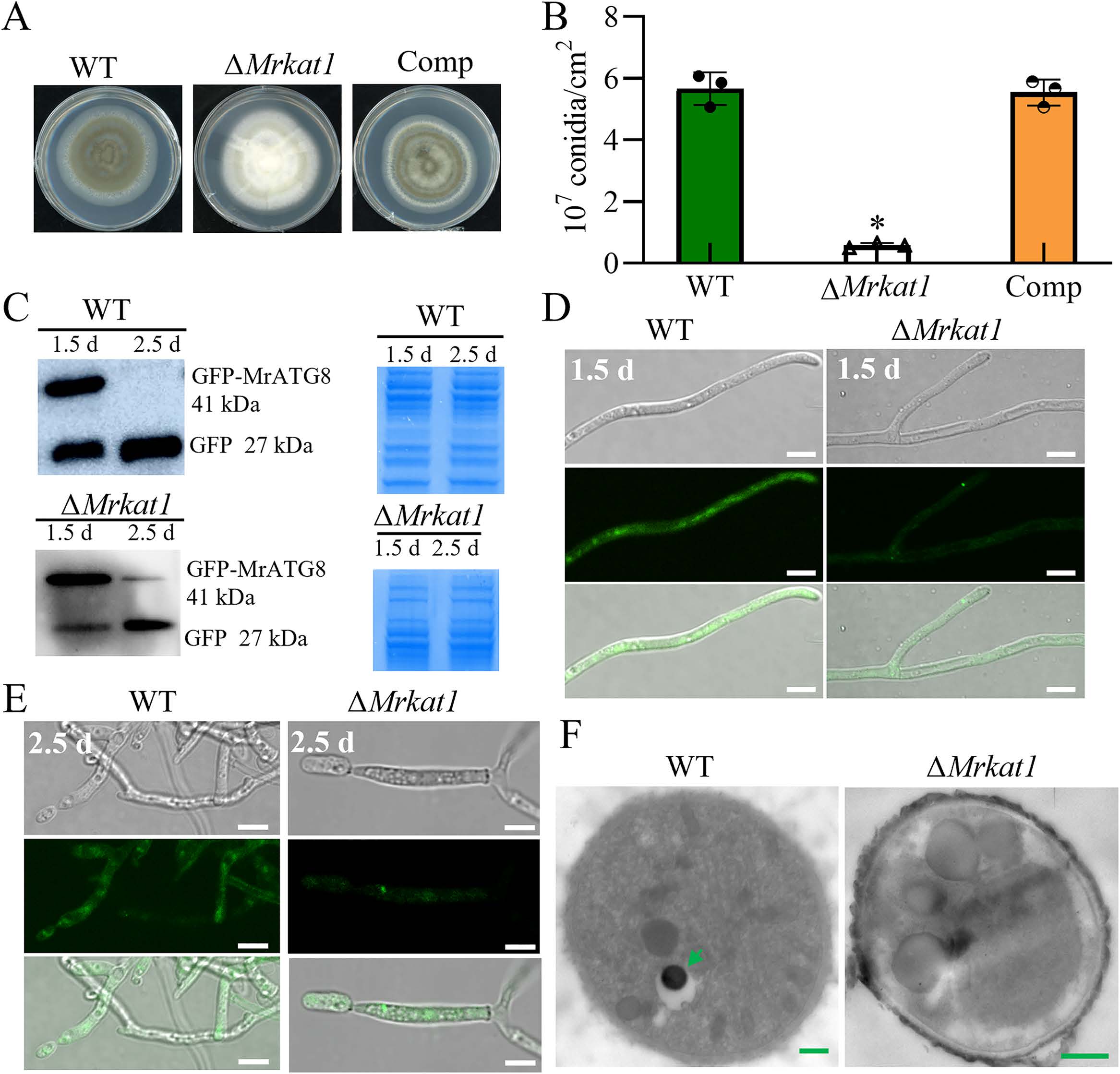
Fig 6. Contribution of MrKAT1 to autophagy and conidial production in M. robertsii. (A and B) Colony phenotyping of WT and mutant strains on PDA media after 14 days of growth at 25 °C. * p < 0.01. (C) Analysis of GFP-MrATG8 degradation in WT and mutant mycelial cells. Immunoblotting was performed with anti-GFP. For each analysis, the parallel-running protein gels were stained as references. * p < 0.01. ( D and E) Cellular location of Autophagosomes during the initial conidia production (2.5 d) and initial mycelial growth (1.5 d) stages. Scale bar: 5 μm. (F) TEM Analysis: This image illustrates the vacuolar localization features of Autophagosomes in the mycelial structures during the conidiation phase for both WT and ΔMrkat1 strains. The autophagic bodies are arrowed in the WT cell. Bar: 1 μm.
Acetylation enhances ATG4 proteolytic activity
ATG4 acetylation and enzyme activity were significantly higher in fungi grown on PDA compared to those grown on SDAY at both 1.5 days and 2.5 days of cultivation (Figs 7A, 7B, S10 and S11). Specifically, ATG4 acetylation and enzyme activity in fungi grown on both PDA and SDAY were elevated at 2.5 days (the stage of early conidiation) compared to those at 1.5 days. Moreover, MrATG4 acetylation levels were markedly increased in the ΔMrsir2–3, whereas they were nearly abolished in the ΔMrkat1 (Fig 7B). Autophagy levels, monitored by ATG8 cleavage, were found to be correlated with the acetylation status of MrATG4 (Figs 7C–7F and S11). Specifically, autophagy levels in the WT were higher at 2.5 days of cultivation compared to 1.5 days. Compare to the WT, autophagy levels of ΔMrkat1 were significantly reduced while substantially increased in the ΔMrsir2–3. These experiments demonstrated that dynamic acetylation of MrATG4, modulated during fungal asexual reproduction, regulates autophagy to facilitate conidiation.
Acetylation of MrATG4 at residues K69 and K77 governs autophagy and conidiation
Given the interactions between MrATG4, MrSIR2–3, and MrKAT1, we hypothesized that MrATG4 is subject to acetylation and deacetylation, modulating its activity in autophagy regulation. Bioinformatics analysis predicted three potential acetylation sites on MrATG4 (K69, K77, K149) (Fig 8A). A triple lysine-to-arginine mutant (3K-R) and three single-site mutants (K69R, K77R, K149R) via site-directed mutagenesis were generated, respectively. Conidial production assays revealed that the Mratg4, K69R and K77R mutants exhibited significantly impaired conidiation compared to WT, while the K149R mutant did not show significant defects (Figs 8B, 8C and S12). Autophagic flux analysis using GFP-MrATG8 corroborated these findings, showing disrupted autophagy in K69R and K77R mutants (Fig 8D). In addition, strains expressing FLAG-tagged MrATG4 (MrATG4-FLAG) and its point mutant strains (ATG4K69R-FLAG, ATG4K77R-FLAG, ATG4K149R-FLAG, ATG4K69,7R7-FLAG) were constructed, and the corresponding proteins were purified using anti-FLAG magnetic beads. Immunoblotting revealed that acetylation levels (detected by an anti-acetyl-lysine antibody) were substantially reduced in the K69R and K77R single mutants, and completely abolished in the K69/77R double mutant. These results demonstrate that lysine residues K69 and K77 of MrATG4 are crucial for its function in autophagy regulation and conidiation. Additionally, MrSIR2–3 and MrKAT1 are potentially associated with these residues, suggesting a possible regulatory interaction and ATG4 may be acetylated at lysine residues 69 and 77.
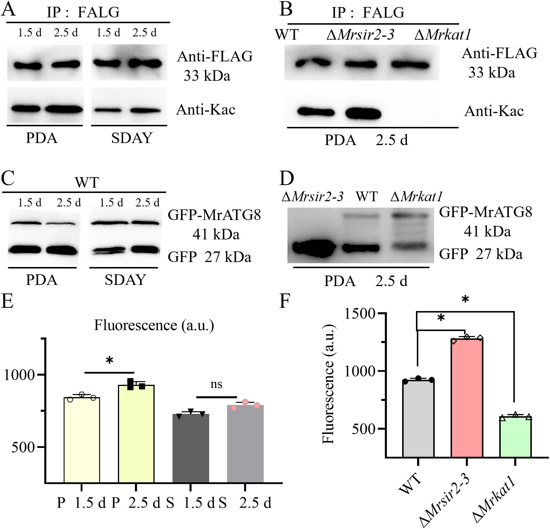
Fig 7. The connection between ATG4 acetylation and ATG4 activity. (A) The WT strain was cultured on PDA or SDAY for 1.5 d, 2.5 d and proteins were immunoprecipitated with antibody to FLAG followed by immunoblotting with antibody to acetylated-lysine. (B) Western blot analyses of the acetylation levels (pan anti-Kac, lower panel) and amount (anti-FLAG, upper panel) of MrATG4-FLAG protein in WT, ΔMrsir2-3 and ΔMrkat1 strains. Proteins were immunoprecipitated with anti-FLAG antibody agarose beads and analyzed using the indicated antibodies. (C) GFP-MrATG8 degradation in WT cells cultured on PDA or SDAY for 1.5 d and 2.5 d was analyzed by immunoblotting using an anti-GFP antibody. For each analysis, the parallel-running protein gels were stained as references (S11 Fig). (D) Analysis of GFP-MrATG8 degradation in WT and mutant cells cultured on PDA for 2.5 d was performed by anti-GFP immunoblotting. For each experiment, duplicate protein gels were run in parallel and stained as loading controls (S11 Fig). (E and F) After treatment as in (C or D), lysates from strains grown under different conditions were assayed for ATG4 activity using the fluorogenic substrate AU4S.
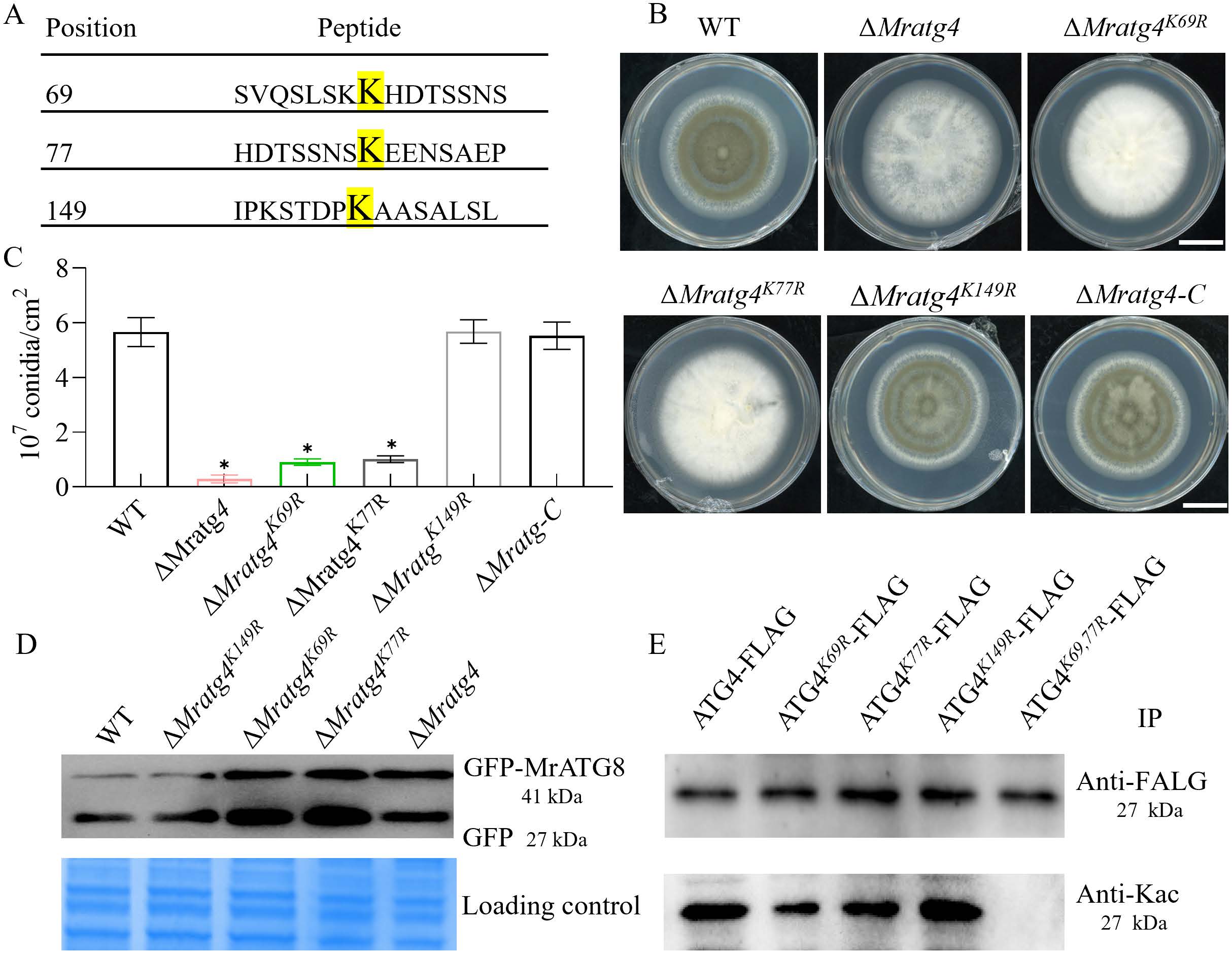
Fig 8. Regulation of autophagy and conidiation by MrATG4 acetylation. (A) Prediction of acetylation sites of MrATG4. Bioinformatics forecast through the prediction website PAIL identified three potential acetylation sites. (B and C) Colony phenotyping of WT and mutant strains on PDA media after 7 days of growth at 25 °C. Quantification of conidial yields by different strains after growth on PDA for 7 days. * p < 0.01. (D) Analysis of GFP-MrATG8 degradation in WT and mutant mycelial cells. Immunoblotting was performed with anti-GFP. For each analysis, the parallel-running protein gels were stained as references. (E) IP assays. Transformants expressing FLAG‑fused MrATG4 and its mutants, including MrATG4K69R-FLAG, MrATG4K77R-FLAG, MrATG4K149R-FLAG, MrATG4K69,77R-FLAG, were cultured on PDA medium for 2.5 days. Proteins were then eluted using anti‑FLAG agarose and analyzed by immunoblotting with an Kac antibody.
Discussions
Autophagy plays a pivotal role in the lifecycle of M. robertsii, particularly in the formation of conidia, which are essential for its efficacy as a biocontrol agent. This study elucidates the intricate regulation of autophagy through the dynamicacetylation and deacetylation of the autophagy-related protein ATG4 by the acetyltransferase MrKAT1 and the deacetylase MrSIR2–3, respectively.
Our findings reveal that MrSIR2–3 acts as a negative regulator of autophagy by deacetylating MrATG4, thereby modulating its activity. Conversely, MrKAT1 promotes autophagy by acetylating MrATG4 possibly at specific lysine residues (K69 and K77). The balance between these opposing enzymatic activities ensures autophagy homeostasis, which is critical for efficient conidiation. Disruption of the balance through the deletion of either MrSIR2–3 or MrKAT1 impaired autophagic flux and conidial production, highlighting the delicate regulation required for optimal fungal development (Fig 9).
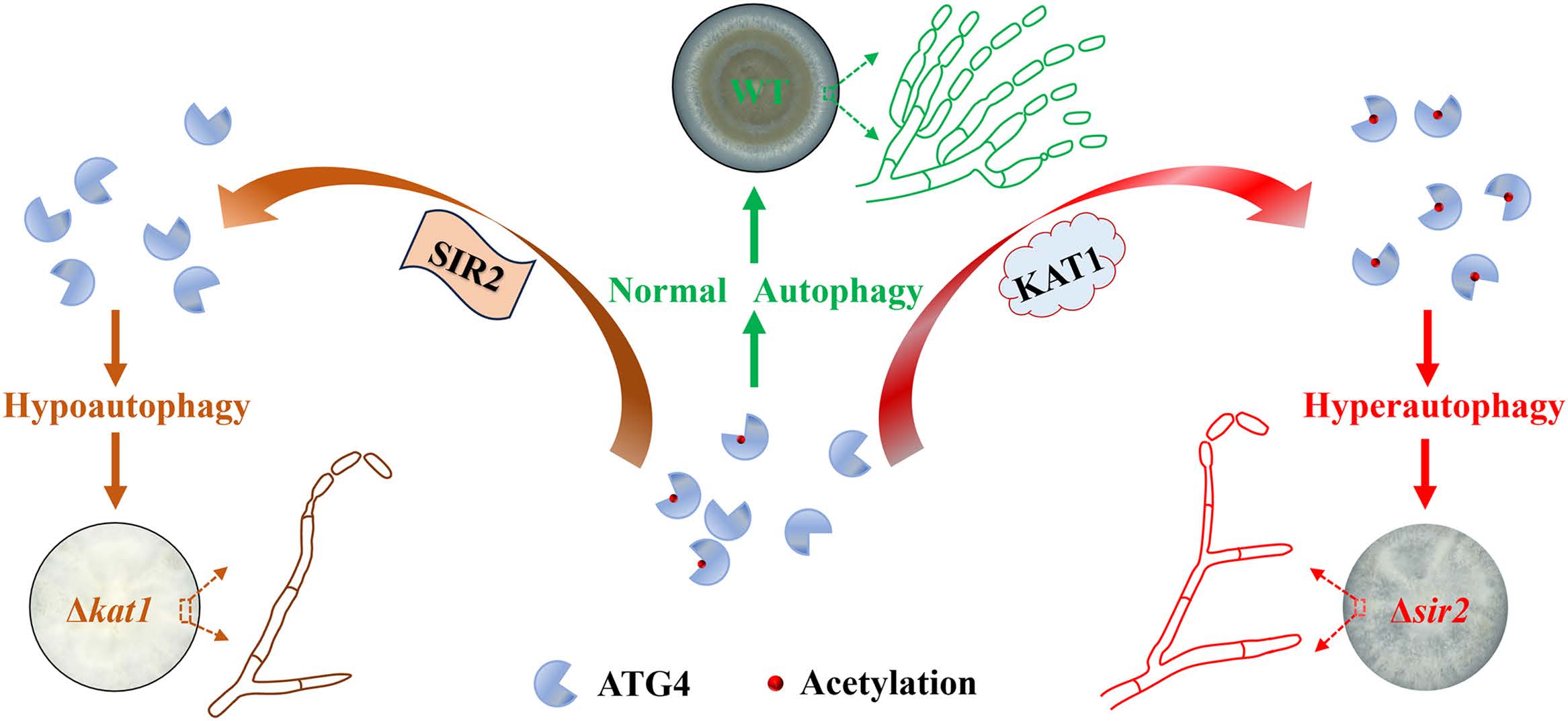
Fig 9. Schematic representation of the ATG4 acetylation regulating autophagy mediated conidiation in fungi. The homeostasis of ATG4 acetylation, mediated by SIR2/KAT1, regulates conidial production by influencing autophagy initiation. Acetyltransferase KAT1 enhances cysteine protease ATG4 activity, promoting hypo-autophagy during initial conidial production. Meanwhile, SIR2-mediated ATG4 deacetylation leads to fungal hyper-autophagy. Both hypo-autophagy and hyper-autophagy result in reduction in conidial production. The proper balance of autophagy ensures normal conidiation.
As previously reported, autophagy is closely associated with the conidiation process in filamentous fungi [27–30]. The SIRT2 enzyme modulates cellular metabolism and ROS stress responses, thereby impacting broader physiological processes, including aging, autophagy, and apoptosis [31–34]. The 90% reduction in conidia production observed in this study could be attributed to either a primary defect in autophagy or a secondary imbalance in metabolic/ROS homeostasis triggered by the absence of Mrsir2–3. Our current experiments indicate that Metarhizium begins to develop sporulation structures after approximately 2.5 days of growth on PDA, which coincides with a notable induction of autophagy. However, the precise conditions that trigger autophagy, and the mechanisms by which MrSIR2–3 and MrKAT1 are activated to initiate conidiation, remain unclear. Further experiments, such as measuring ATP/AMP and NAD ⁺ /NADH ratios or conducting rescue experiments with an ATP-generating carbon source, are necessary to determine whether the sporulation defect is specifically linked to autophagy or secondary metabolic/ROS stress. These additional studies will provide more insights into the complex regulatory networks governing conidiation and stress responses in Metarhizium.
ATG4, as a key role in the regulation of autophagy by cutting and recycling ATG8 [13], has a significant impact on fungal autophagy and conidiation in fungi [1,5,6,28]. In our work, ΔMratg4 induced the loss of autophagy and conidiation, aligning with earlier reports [4]. Furthermore, we discovered that MrATG4 interacted with MrSIR2–3 and MrKAT1,respectively. This interaction is in line with findings in mammals, where the deacetylation of ATG4B was established as a crucial step in initiating autophagy during starvation [18]. Our results on MrATG4 interaction and acetylation represent the first report in fungi, suggesting that ATG4 is regulated through multiple mechanisms and offering new insights into its acetylation control in fungi.