接第一篇:
3.2. Taxonomy
3.2.1. Samsoniella pseudogunniiW.H. Chen, Y.F. Han, J.D. Liang, and Z.Q. Liang, sp. nov.
MycoBank No.: MB840999
Etymology: referring to similar morphology with Keithomyces neogunnii.
Holotype: CHINA, Guizhou, Guiyang, Tongmuling (N26°23’, E106°40’). On a larva (Lepidoptera), 1 April 2019, Wanhao Chen, GZAC GY40720 (holotype), ex-type living cultures, GY407201, GY407202.
Description: Colonies on PDA, 4.1–4.3 cm diam. after 14 d at 25℃, white, consisting of a basal felt and cottony, floccose hyphal overgrowth, reverse yellowish. Prostrate hyphae smooth, septate, hyaline, 1.0–1.3 μm diam. Erect conidiophores usually arises from aerial hyphae. Phialides are solitary or in whorls of two to nine. Phialides 6.8–11.0 × 2.2–2.4 μm, with a cylindrical basal portion, tapering into a short distinct neck. Conidia in chains, hyaline, fusiform, one-celled, 2.8–3.2 × 1.7–2.1 μm. Chlamydospores and sexual state were not observed. Sizes and shapes of phialides and conidia are similar in culture and on natural substratum.
Known distribution: Tongmuling, Guiyang, Guizhou Province, China.
Notes: Samsoniella pseudogunnii was identified as belonging to Samsoniella based on the phylogenetic analyses (Figure 2) and has a close relationship with S. coleopterorum. However, Samsoniella pseudogunnii (Figure 3) has longer phialide, larger conidia, and its larva host belongs to the order Lepidoptera
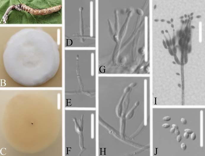
Figure 3. Samsoniella pseudogunnii (A) infected larva (Lepidoptera) (B,C) culture plate, showing the front (B) and the reverse (C) of the colony, cultured on PDA medium (D–I) phialides solitary, conidia adhering ellipsoidal slimy head and conidia (J) conidia. Scale bars: 10 mm (B,C), 10 μm (D–J).
3.2.2. Samsoniella PupicolaW.H. Chen, Y.F. Han, J.D. Liang, and Z.Q. Liang, sp. nov.
MycoBank No.: MB841003
Etymology: referring to its pupa-inhabitor.
Holotype: CHINA, Guizhou, Qiannan Buyi and Miao Autonomous Prefecture, Duyun City (26°21'24.71" N, 107°22'48.22" E). On a pupa (Lepidoptera), 1 October 2019, Wanhao Chen, GZAC DY10168 (holotype), ex-type living cultures, DY101681, DY101682.
Description: Colonies on PDA, 2.3–2.4 cm diam. after 14 d at 25℃, white, consisting of a basal felt and cottony, floccose hyphal overgrowth, reverse yellowish. Prostrate hyphae smooth, septate, hyaline, 1.2–2.2 μm diam. Erect conidiophores usually arise from aerial hyphae. Phialides are solitary or in whorls of two to nine. Phialides 7.0–9.2 × 2.5–3.3 μm, with a cylindrical basal portion, tapering into a short distinct neck. Conidia in chains, hyaline, fusiform, one-celled, 2.5–3.3 × 2.2–2.6 μm. Chlamydospores and sexual state were not observed. Sizes and shapes of phialides and conidia are similar in culture and on natural substratum.
Known distribution: Duyun City, Qiannan Buyi and Miao Autonomous Prefecture, Guizhou Province, China.
Additional specimens examined: CHINA, Guizhou, Qiandongnan Miao and Dong Autonomous Prefecture, Rongjiang County (26°01'58.70" N, 108°24'48.06" E), on a lepidopteran pupa, 1 October 2018, W.H. Chen, GZAC DL1014.
Notes: Samsoniella pupicola was identified as belonging to Samsoniella, based on the phylogenetic analyses (Figure 2) and has a close relationship with S. alboaurantium, S. alpina, and S. cardinalis. However, S. pupicola (Figure 4) is distinguished from S. alboaurantium by having larger fusiform conidia, distinguished from S. alpina by having white colony and fusiform conidia, and distinguished from S. cardinalis by having shorter phialides.
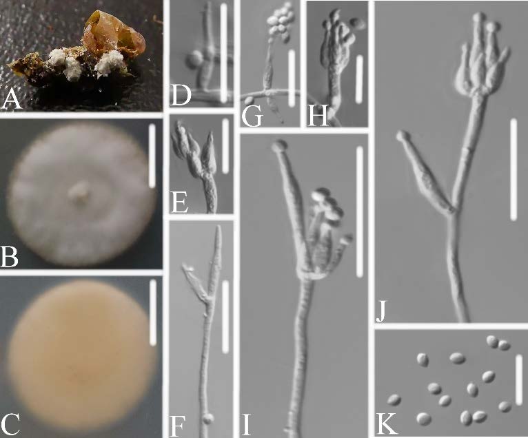
Figure 4. Samsoniella pupicola (A) infected pupa (Lepidoptera) (B,C) culture plate, showing the front (B) and the reverse (C) of the colony, cultured on PDA medium (D–J) phialides solitary, conidia adhering ellipsoidal slimy head and conidia (K) conidia. Scale bars: 10 mm (B,C), 10 μm (D–K).
3.2.3. Samsoniella aurantia Mongkols., Noisrip., Thanakitp., Spatafora, and Luangsa-ard,Mycologia 110(1): 249
Description: Colonies on PDA, 3.7–4.2 cm diam. after 14 d at 25°C, white, consisting of a basal felt and cottony, floccose hyphal overgrowth, pale green and pale pink in the middle of the colony, reverse yellowish and pale brown in the middle. Prostrate hyphae smooth, septate, hyaline, 1.3–2.6 μm diam. Erect conidiophores usually arise from aerial hyphae. Phialides are solitary or in whorls of two to ten. Phialides 3.6–7.7 × 1.3–1.6 μm, with a cylindrical basal portion, tapering into a short distinct neck. Conidia in chains, hyaline, fusiform, occasionally cylindrical, one-celled, 2.6–3.9 × 1.7–2.2 μm. Chlamydospores and sexual state were not observed. Sizes and shapes of phialides and conidia are similar in culture and on natural substratum.
Specimens examined: CHINA, Guizhou, Qiannan Buyi and Miao Autonomous Prefecture,Duyun City (26°21'24.71" N, 107°22'48.22" E). On a pupa (Lepidoptera), 1 October 2019,Wanhao Chen, GZAC DY1095, living cultures, DY10951, DY10952.
Note: DY10951 and DY10952 strains were identified as belonging to Samsoniella, based on the phylogenetic analyses (Figure 2), and clustered with Samsoniella aurantia in a clade. The characteristics of DY10951 and DY10952 (Figure 5) strains are similar to that of S. aurantia, which had fusiform conidia (2–4 × 1–2 μm) and larger phialide (5–13 × 2–3 μm). Besides, the pairwise dissimilarities of ITS sequences show no difference within 554 bp between DY10951 and S. aurantia. Thus, molecular phylogenetic results and morphologically based conclusions support the idea that DY10951 and DY10952 strains were S. aurantia.
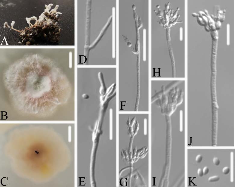
Figure 5. Samsoniella aurantia (A) infected pupa (Lepidoptera) (B,C) culture plate, showing the front (B) and the reverse (C) of the colony, cultured on PDA medium (D–J) phialides solitary, conidia adhering ellipsoidal slimy head and conidia (K) conidia. Scale bars: 10 mm (B,C), 10 μm (D–K).
3.2.4. Samsoniella hepiali (Q.T. Chen, and R.Q. Dai ex R.Q. Dai, X.M. Li, A.J. Shao, Shu F. Lin, J.L. Lan, Wei H. Chen, and C.Y. Shen) H. Yu, R.Q. Dai, Y.B. Wang, Y. Wang, and Zhu L. Yang, Fungal Diversity 103: 31
Description: Colonies on PDA, 5.8–5.9 cm diam. after 14 d at 25°C, white, consisting of a basal felt and cottony, floccose hyphal overgrowth, reverse yellowish. Prostrate hyphae
smooth, septate, hyaline, 1.1–1.8 μm diam. Erect conidiophores usually arise from aerial hyphae. Phialides are solitary or in whorls of two to eight. Phialides 6.0–7.8 × 1.5–1.8 μm, with a cylindrical basal portion, tapering into a short distinct neck. Conidia in chains, hyaline, fusiform, one-celled, 2.1–2.5 × 0.9–1.6 μm. Chlamydospores and sexual state were not observed. Sizes and shapes of phialides and conidia are similar in culture and on natural substratum.
Specimens examined: CHINA, Guizhou, Tongren City, Yinjiang (N 27°55’17.1”, E 108°41’25.2”), on an ant, 1 October 2019, Wanhao Chen, GZAC YJ0617, DY1044, living cultures, YJ06171, YJ06172.
Note: YJ06171 and YJ06172 strains were identified as belonging to Samsoniella, based on the phylogenetic analyses (Figure 2), and clustered with Samsoniella hepiali in a clade. The characteristics of YJ06171 and YJ06172 (Figure 6) strains were very closely linked with S. hepiali, which had fusiform or oval conidia (1.8–3.3 × 1.4–2.2 μm) and larger phialide (3.5–13.6 × 1.3–2.1 μm). Besides, the pairwise dissimilarities of LSU sequences show no difference within 677 bp between YJ06171 and S. hepiali. Thus, molecular phylogenetic results and morphologically based conclusions supported the idea that YJ06171 and YJ06172 strains were S. hepiali.
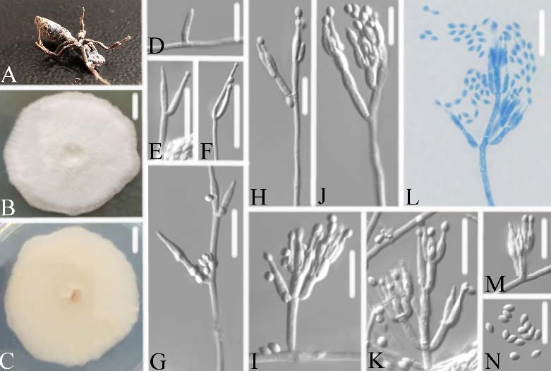
Figure 6. Samsoniella hepiali (A) infected ant (Formicidae) (B,C) culture plate, showing the front (B) and the reverse (C) of the colony, cultured on PDA medium (D–J) phialides solitary, conidia adhering ellipsoidal slimy head and conidia (N) conidia. Scale bars: 10 mm (B,C), 10 μm (D–N).
4. Discussion
The taxonomic delimitation of Isaria was originally based on morphological characteristics.However, Isaria shares many morphological characters with other genera in Hypocreales, which has resulted in a turbulent taxonomic history [10,29]. D’Alessandro et al. [30] noted that the morphological characteristics used to classify the genus Isaria frequently do not resolve new isolates into clearly defined species and need additional molecular markers in phylogenetic analyses. In the present study, Isaria-like strains collected from Guizhou Province, China, and previously identified by morphological characteristics, were reanalyzed using multi-gene (ITS, LSU, RPB1, RPB2, TEF) phylogenetic methodology. We proposed two new species of Samsoniella in this study.
The species Isaria farinosa is a well-known entomopathogenic fungi with worldwide distribution and a wide host range [31]. Kepler et al. [9] transferred Isaria farinosa to the genus Cordyceps as C. farinosa (Holmsk.) Kepler, B. Shrestha, and Spatafora based on a phylogenetic analysis of the CBS 111113 strain. We analyzed several strains of Isaria farinosa in the present study. Some properly belonged in the genus Samsoniella. CEP 004, CEP 005, CEP 029, YJ06171, and YJ06172 strains were identified as S. hepiali. Strains DY10951 and DY10952 were identified as S. aurantia. OSC 111005 and OSC 111006 strains were identified as new species but are absent in delineating morphological characteristics. Our results reveal cryptic diversity present in Isaria farinosa (now treated as Cordyceps farinosa) and illustrated that more attention should be paid on cryptic intraspecific diversity across different fungi isolates and genotypes.
The genus Samsoniella was established for the typical species S. inthanonensis Mongkolsamrit, Noisripoom, Thanakitpipattana, Spatafora, and Luangsa-ard, and two other species (S. alboaurantia (G. Sm.) Mongkols., Noisrip., Thanakitp., Spatafora, and Luangsa-ard and S. aurantia Mongkols., Noisrip., Thanakitp., Spatafora, and Luangsa-ard) [10]. Samsoniella species all have Isaria-like morphological characteristics, and cluster in an independent clade with close relationship to the genus Akanthomyces. The species S. alboaurantia was established based on two strains, CBS 240.32 and CBS 262.58, which previously belonged to Isaria farinosa (originally designated Paecilomyces farinosus (Holmsk.) A.H.S. Br. and G. Sm.) [10]. Lin et al. [32] revised the taxonomy of some Isaria-like strains originally identified as Isaria farinosa by morphological characteristics using multi-gene phylogenetic analysis. All the strains were identified as Samsoniella hepiali (Q.T. Chen and R.Q. Dai ex R.Q. Dai, X.M. Li, A.J. Shao, Shu F. Lin, J.L. Lan,Wei H. Chen, and C.Y. Shen) H. Yu, R.Q. Dai, Y.B.Wang, Y.Wang, and Zhu L. Yang. In the present study, YJ06171 and YJ06172 strains were also identified as Samsoniella hepiali. Our results revealed that more isolates and genotypes, originally designated as Isaria, will need to be transferred to Samsoniella.
Samsoniella hepiali (otherwise known as Paecilomyces hepiali) is isolated from a field collection of natural Ophiocordyceps sinensis insect–fungi complex [33], and is widely used as a medicinal and edible cordycipitoid fungus, creating a great economic value [20]. Lin et al. [32] reported six isolates of Samsoniella hepiali from Anhui Province, China, which were isolated from leafhopper, larva, and cicada. CEP 004, CEP 005, CEP 029 strains from Buenos Aires, Argentina, were isolated from whitefly and soil [30]. YJ06171 and YJ06172 strains from Guizhou Province, China were isolated from ant. It is interesting that Samsoniella hepiali and its hosts are widely distributed in China and Argentina. This result will help us to assess the extent and distribution of genetic diversity of Samsoniella hepiali on a large scale, understand its biology and demographic history, and guide biodiversity conservation programs.
Author Contributions: Resources,W.C., J.L. and X.R.; data curation,W.C.; writing—original draft preparation, W.C., J.L., X.R.; writing—review and editing, J.L., Y.H.; review and editing, J.Z., Z.L.; funding acquisition, W.C., J.L., X.R., J.Z., Y.H. All authors have read and agreed to the published version of the manuscript.
Funding: This research was funded by National Natural Science Foundation of China (31860002, 32060011), High-level Innovative Talents Training Object in Guizhou Province (Qiankehepingtairencai [2020]6005), Science and Technology Foundation of Guizhou Province (Qiankehejichu [2020]1Y060), Program of Innovative Scientific and technological Talent Team of Guizhou Province (2020-5010), Guizhou Science and Technology Support Project (Qiankehezhicheng [2019]2776), The Youth Science and Technology Talent Growth project from Guizhou Provincial Department of Education ([2018]389). Institutional Review Board Statement: Not applicable.
Informed Consent Statement: Not applicable.
Data Availability Statement: Not applicable.
Acknowledgments: We thank Steven M. Thompson for editing the English text of a draft of this manuscript.
Conflicts of Interest: The authors declare no conflict of interest.
References
1. Fries, E.M. Systema Mycologicum; Ex Officina Berlingiana: Lund & Greifswald, Sweden, 1821; Volume 1, pp. 1–726.
2. Brown, A.H.; Smith, G. The genus Paecilomyces Bainier and its perfect stage Byssochlamys Westling. Trans. Br. Mycol. Soc. 1957, 40, 17–89. [CrossRef]
3. De Hoog, G.S. The genera Beauveria, Isaria, Tritirachium and Acrodontium Gen. Nov; Centraalbureau voor Schimmelcultures: Utrecht, The Netherlands, 1972; pp. 1–41.
4. Samson, R.A. Paecilomyces and some allied Hyphomycetes; Centraalbureau voor Schimmelcultures: Utrecht, The Netherlands, 1974; Volume 6, pp. 1–119.
5. Hodge, K.T.; Gams, W.; Samson, R.A.; Korf, R.P.; Seifert, K.A. Lectotypification and status of Isaria Pers.: Fr. Taxon 2005, 54, 485–489. [CrossRef]
6. Luangsa-ard, J.J.; Hywel-Jones, N.L.; Samson, R.A. The order level polyphyletic nature of Paecilomyces sensu lato as revealed through 18S-generated rRNA phylogeny. Mycologia 2004, 96, 773–780. [CrossRef] [PubMed]
7. Luangsa-Ard, J.J.; Hywel-Jones, N.L.; Manoch, L.; Samson, R.A. On the relationships of Paecilomyces sect. Isarioidea species. Mycol. Res. 2005, 109, 581–589. [CrossRef]
8. Gams, W.; Hodge, K.T.; Samson, R.A.; Korf, R.P.; Seifert, K.A. (1684) Proposal to conserve the name Isaria (anamorphic fungi) with a conserved type. Taxon 2005, 54, 537. [CrossRef]
9. Kepler, R.M.; Luangsa-ard, J.J.; Hywel-Jones, N.L.; Quandt, A.; Sung, G.-H.; Rehner, S.A.; Aime, M.C.; Henkel, T.W.; Sanjuan, T.; Zare, R.; et al. A phylogenetically-based nomenclature for Cordycipitaceae (Hypocreales). IMA Fungus 2017, 8, 335–353.[CrossRef]
10. Mongkolsamrit, S.; Noisripoom, W.; Thanakitpipattana, D.; Wutikhun, T.; Spatafora, J.W.; Luangsa-ard, J. Disentangling cryptic species with isaria-like morphs in Cordycipitaceae. Mycologia 2018, 110, 230–257. [CrossRef]
11. Chen, W.-H.; Liu, C.; Han, Y.-F.; Liang, J.-D.; Liang, Z.-Q. Akanthomyces araneogenum, a new Isaria-like araneogenous species. Phytotaxa 2018, 379, 66–72. [CrossRef]
12. Chen,W.H.; Han, Y.F.; Liang, J.D.; Tian,W.Y.; Liang, Z.Q. Morphological and phylogenetic characterizations reveal three new species of Samsoniella (Cordycipitaceae, Hypocreales) from Guizhou, China. MycoKeys 2020, 74, 1–15. [CrossRef] [PubMed]
13. Chen, W.-H.; Liu, C.; Han, Y.-F.; Liang, J.-D.; Tian, W.-Y.; Liang, Z.-Q. Three novel insect-associated species of Simplicillium (Cordycipitaceae, Hypocreales) from Southwest China. MycoKeys 2019, 58, 83–102. [CrossRef] [PubMed]
14. Zou, X.; Liu, A.; Liang, Z.; Han, Y.; Yang, M. Hirsutella liboensis, a new entomopathogenic species affecting Cossidae (Lepidoptera) in China. Mycotaxon 2010, 111, 39–44. [CrossRef]
15. Liang, J.D.; Han, Y.F.; Zhang, J.W.; Du,W.; Liang, Z.Q.; Li, Z.Z. Optimal culture conditions for keratinase production by a novel thermophilic Myceliophthora thermophila strain GZUIFR-H49-1. J. Appl. Microbiol. 2011, 110, 871–880. [CrossRef]
16. White, T.; Bruns, T.; Lee, S.; Taylor, J. Amplification and Direct Sequencing of Fungal Ribosomal RNA Genes for Phylogenetics. In PCR Protocols: A Guide to Methods and Applications; Innis, M.A., Gelfand, D.H., Sninsky, J.J., White, T.J., Eds.; Academic Press: New York, NY, USA, 1990; pp. 315–322. [CrossRef]
17. Rakotonirainy, M.; Cariou, M.; Brygoo, Y.; Riba, G. Phylogenetic relationships within the genus Metarhizium based on 28S rRNA sequences and isozyme comparison. Mycol. Res. 1994, 98, 225–230. [CrossRef]
18. Castlebury, L.A.; Rossman, A.Y.; Sung, G.-H.; Hyten, A.S.; Spatafora, J.W. Multigene phylogeny reveals new lineage for Stachybotrys chartarum, the indoor air fungus. Mycol. Res. 2004, 108, 864–872. [CrossRef] [PubMed]
19. Van den Brink, J.; Samson, R.A.; Hagen, F.; Boekhout, T.; de Vries, R.P. Phylogeny of the industrial relevant, thermophilic genera Myceliophthora and Corynascus. Fungal Divers. 2012, 52, 197–207. [CrossRef]
20. Wang, Y.-B.; Wang, Y.; Fan, Q.; Duan, D.-E.; Zhang, G.-D.; Dai, R.-Q.; Dai, Y.-D.; Zeng, W.-B.; Chen, Z.-H.; Li, D.-D.; et al. Multigene phylogeny of the family Cordycipitaceae (Hypocreales): New taxa and the new systematic position of the Chinese cordycipitoid fungus Paecilomyces hepiali. Fungal Divers. 2020, 103, 1–46. [CrossRef]
21. Katoh, K.; Standley, D.M. MAFFT multiple sequence alignment software version 7: Improvements in performance and usability. Mol. Biol. Evol. 2013, 30, 772–780. [CrossRef] [PubMed]
22. Tamura, K.; Stecher, G.; Peterson, D.; Filipski, A.; Kumar, S. MEGA6: Molecular evolutionary genetics analysis version 6.0. Mol. Biol. Evol. 2013, 30, 2725–2729. [CrossRef] [PubMed]
23. Vaidya, G.; Lohman, D.J.; Meier, R. SequenceMatrix: Concatenation software for the fast assembly of multi-gene datasets with character set and codon information. Cladistics 2011, 27, 171–180. [CrossRef]
24. Kalyaanamoorthy, S.; Minh, B.Q.; Wong, T.; Von Haeseler, A.; Jermiin, L.S. ModelFinder: Fast model selection for accurate phylogenetic estimates. Nat. Methods 2017, 14, 587–589. [CrossRef]
25. Zhang, D.; Gao, F.; Jakovli´c, I.; Zou, H.; Zhang, J.; Li, W.X.; Wang, G.T. PhyloSuite: An integrated and scalable desktop platform for streamlined molecular sequence data management and evolutionary phylogenetics studies. Mol. Ecol. Resour. 2020, 20, 348–355. [CrossRef]
26. Ronquist, F.; Teslenko, M.; Van Der Mark, P.; Ayres, D.L.; Darling, A.; Hoehna, S.; Larget, B.; Liu, L.; Suchard, M.A.; Huelsenbeck,J.P. MrBayes 3.2: Efficient Bayesian Phylogenetic Inference and Model Choice Across a Large Model Space. Syst. Biol. 2012, 61,539–542. [CrossRef] [PubMed]
27. Drummond, A.J.; Rambaut, A. BEAST: Bayesian evolutionary analysis by sampling trees. BMC Evol. Biol. 2007, 7, 214. [CrossRef]
28. Silvestro, D.; Michalak, I. raxmlGUI: A graphical front-end for RAxML. Org. Divers. Evol. 2012, 12, 335–337. [CrossRef]
29. Sung, G.-H.; Hywel-Jones, N.L.; Sung, J.-M.; Luangsa-ard, J.J.; Shrestha, B.; Spatafora, J.W. Phylogenetic classification of Cordyceps and the clavicipitaceous fungi. Stud. Mycol. 2007, 57, 5–59. [CrossRef]
30. D’Alessandro, C.P.; Jones, L.R.; Humber, R.A.; López Lastra, C.C.; Sosa-Gomez, D.R. Characterization and phylogeny of Isaria spp. strains (Ascomycota: Hypocreales) using ITS 1-5.8 S-ITS 2 and elongation factor 1-alpha sequences. J. Basic Microbiol. 2014, 54, S21–S31. [CrossRef]
31. Zimmermann, G. The entomopathogenic fungi Isaria farinosa (formerly Paecilomyces farinosus) and the Isaria fumosorosea species complex (formerly Paecilomyces fumosoroseus): Biology, ecology and use in biological control. Biocontrol Sci. Technol. 2008, 18, 865–901. [CrossRef]
32. Lin, Y.; Liu, Y.J.;Wang, T.; Chen, M.J. Revision of taxonomic status of several Isaria-like strains. J. Microbiol. China 2021. (In Chinese) [CrossRef]
33. Dai, R.-Q.; Shen, C.-Y.; Li, X.-M.; Lan, J.-L.; Lin, S.-F.; Shao, A.-J. Response to neotypification of Paecilomyces hepiali (Hypocreales) (Wang & al., 2015). Taxon 2018, 67, 784–786. [CrossRef]