Research Article
Thuan Duc Lao#, Hanh Van Trinh#, Loi Vuong, Luyen Tien Vu, Thuy Ai Huyen Le,Hiep Minh Dinh, Nguyen Binh Truong*
Molecular record for the first authentication of Isaria cicadae from Vietnam
https://doi.org/10.1515/biol-2021-0074
received February 18, 2020; accepted June 19, 2021
Abstract: The entomopathogenic fungus T011, parasitizing on nymph of Cicada, collected in the coffee garden in Dak Lak Province, Vietnam, was preliminarily morphologically identified as Isaria cicadae, belonged to order Hypocreales and family Clavicipitaceae. To ensure the authenticity of T011, phylogenetic analysis of the concatenated set of multiple genes including ITS, nrLSU, nrSSU,Rpb1, and Tef1 was applied to support the identification. Genomic DNA was isolated from dried sample T011. The PCR assay sequencing was applied to amplify ITS, nrLSU,nrSSU, Rpb1, and Tef1 gene. For phylogenetic analysis,the concatenated data of both target gens were constructed with MEGAX with a 1,000 replicate bootstrap based on the neighbor-joining, maximum likelihood, maximum parsimony method. As the result, the concatenated data containing 62 sequences belonged to order Hypocreales, families Clavicipitaceae, and 2 outgroup sequences belonged to order Hypocreales, genus Verticillium. The phylogenetic analysis results indicated that T011 was accepted at subclade Cordyceps and significantly formed the monophyletic group with referent Cordyceps cicadae (Telemorph of Isaria cicadae) with high bootstrap value. The phylogenetically analyzed result was strongly supported by our morphological analysis described as the Isaria cicadae. In summary, phylogenetic analyses based on the concatenated dataset were successfully applied to strengthen the identification of T011 as Isaria cicadae.
Keywords: nuclear small ribosomal subunit, nuclear large ribosomal subunit, Isaria cicadae, phylogeny
# Equal contributors
* Corresponding author: Nguyen Binh Truong, Faculty of Biology,Dalat University, Lam Dong, Vietnam, e-mail: nguyentb@dlu.edu.vn
Thuan Duc Lao, Luyen Tien Vu, Thuy Ai Huyen Le: Department of Pharmaceutical andMedical Biotechnology, Faculty of Biotechnology,Ho Chi Minh City Open University, Ho Chi Minh City, Vietnam
Hanh Van Trinh: University of Science, VNU-HCM, Ho Chi Minh City,Vietnam
Loi Vuong: University of Science, VNU-HCM, Ho Chi Minh City,Vietnam; Institute of Applied Technology, Thu Dau Mot University,Binh Duong, Vietnam
Hiep Minh Dinh: Department of Agriculture and Rural Development of Ho Chi Minh City, Ho Chi Minh City, Vietnam
1 Introduction
Isaria cicadae Miq., Bull. Sci. phys. nat. néerl.: 86 (1838) (Mycobank: MB#204858), also known Cordyceps cicadae(Miq.) Massee (1895) (Mycobank: MB#311793), is the entomopathogenic fungi capable of parasitizing on cicada nymph, belongs to the order Hypocreales, and the family Clavicipitaceae [4,5]. C. cicadae usually distribute in many regions of the world with temperatures ranging from 18 to 24°C, relative humidity of >80°C, and grows vertically on the sunny slopes at an attitude of 700–950m[3]. The distribution of C. cicadae is recorded in China(Province of Yunan, Sichuan, Guizhou, Jiangsu, Guangdong,Hunan, Hubei, etc.), Korea (Jeju Island), and Japan(South of Fukushima). Furthermore, C. cicadae is also seen in Thailand, North America, and Europe [3–24].
Due to their numerous bioactivities, I. cicadae, as well as C. cicadae, is considered the most valued traditional Chinese medicine. Its medicinal bioactive components,such as adenosine, cordycepin, ergosterol, etc.,which have been used to relieve exhaustion remedy, treat numerous diseases, such as antitumor activities, and food source, have been recorded [3,19–22]. To obtain precious valued herbal medicine, the exploration and collection of local I. cicadae (C. cicadae) play an important role to apply for further medicinal applications. During our expedition to validate the fungal diversity in Ea Knop Town – Ea Kar District (Latitude: 12°34′26″N–13°02′09″N; Longitude: 108°22′08″E–108°43′2″E) located in Dak Lak Province, we collected the sample T011, parasitizing on the nymph of Cicada, which was classified and confirmed by the specialist on the entomologist, Faculty of Biotechnology, Ho Chi Minh City Open University, Ho Chi Minh City, Vietnam. In this paper, to ensure the origin and authenticity of T011 as I. cicadae, we conducted the morphology analysis and molecular phylogenetic analysis of the concatenated set genes including ITS, nrLSU,nrSSU, Rpb1, and Tef1.
2 Materials and methods
2.1 Sample collection
The specimen T011, parasitizing on the nymph of Cicada,was collected in the coffee garden in Ea Knop Town – Ea Kar District, Dak Lak Province on the morning of June 24,2018. In the laboratory, the specimen was conditioned to be dried at 60°C and stored for further analysis.
2.2 Morphology analysis
Macroscopic characteristics of the fresh body were carefully observed in the many macroscopic characteristics. For the microscopic analysis, a bunch of conidiogenous cells was cut into small species, then, soaked in the water for about 3 min. A sample of the synemata containing the conidiogenous cells was immersed in distilled water for 3 min. Asexual spores were removed using a clean brush.The fertile part was then analyzed under a microscope.Conidia size was recorded. According to the identification of conidia, phialides, and colony coloration, the isolate cultures were grown on YMG media, composed of 4 g/L Yeast extract, 10 g/L Malt extract, 4 g/L Glucose, incubated at 20°C within a period of 20 days.
2.3 DNA extraction, PCR amplification,target gene sequencing
Genomic DNA was extracted from dried material by using the phenol/chloroform method (pH = 8). The dried material was added to a lysis buffer (2.0%SDS, Tris-HCl pH 8.0,150 mM NaCl, 10 mM EDTA, 0.1 mg/mL Proteinase K).During the incubation at 65°C for overnight, it was mixed thoroughly by inverting the tube several times. Then, the supernatant was collected by centrifugation. About 700 μL of phenol/chloroform/isoamyl alcohol at a ratio of 25:24:1 was added and centrifuged. The upper solution was collected, precipitated with absolute ethanol, and washed with 70% ethanol. DNA concentration was identified by using OD260. Finally, isolated genomic DNA was stored in Tris-EDTA buffer at −20°C for further studies.
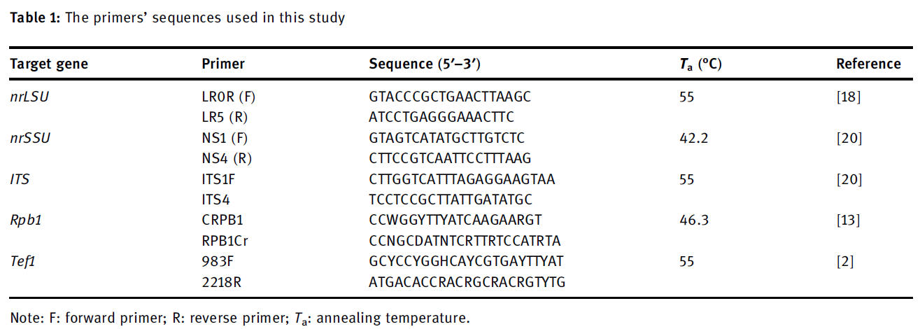
The primer pairs used to amplify ITS, nrLSU, nrSSU,Rpb1, and Tef1 region were shown in Table 1. The final volume for PCR was done in the total of 15 μL with the thermal program: 1 cycle for 95°C for 5 min; 40 cycles of 95°C for 30 s, X°C for 30 s, 72°C for 2min; 1 cycle for 72°C for 5min (Note: X°C is the annealing temperatures for each target gene, shown in Table 1). About 5 μL aliquots of amplification products were electrophoresed on a 2.0% agarose gel and visualized in a UV transilluminator. The amplified product was sequenced at Nam Khoa (Vietnam) company.
2.4 Taxa and ITS, nrLSU, nrSSU, Rpb1, and Tef1 sequences collection, DNA proofreading, and phylogeny analysis
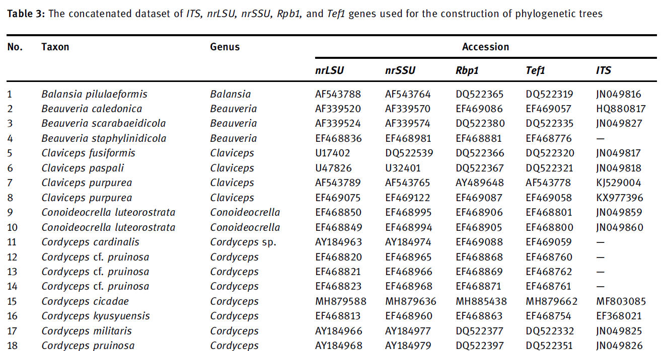
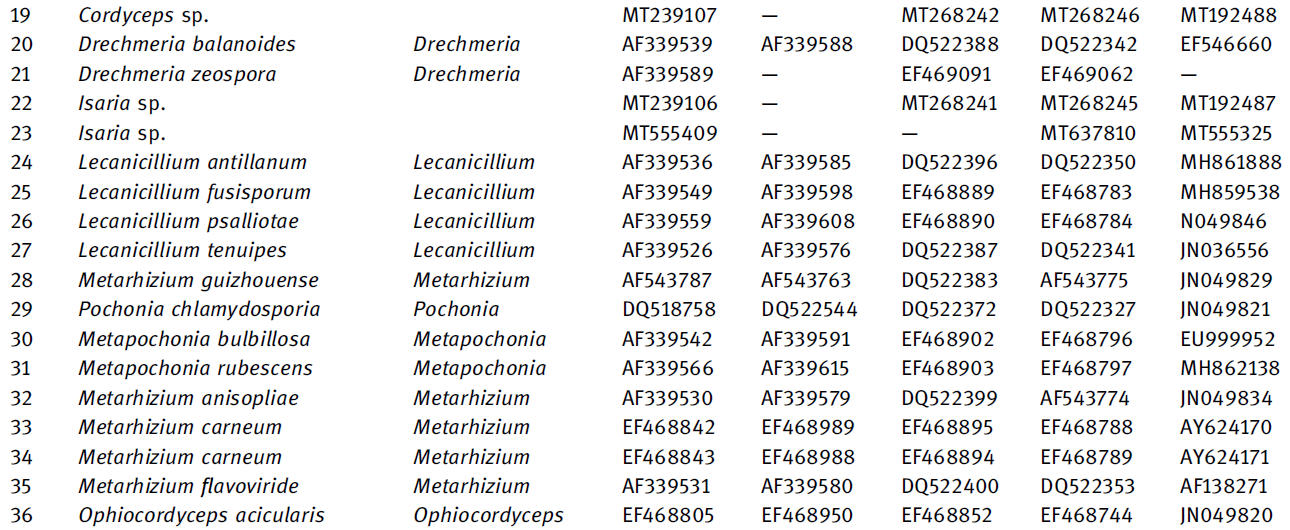
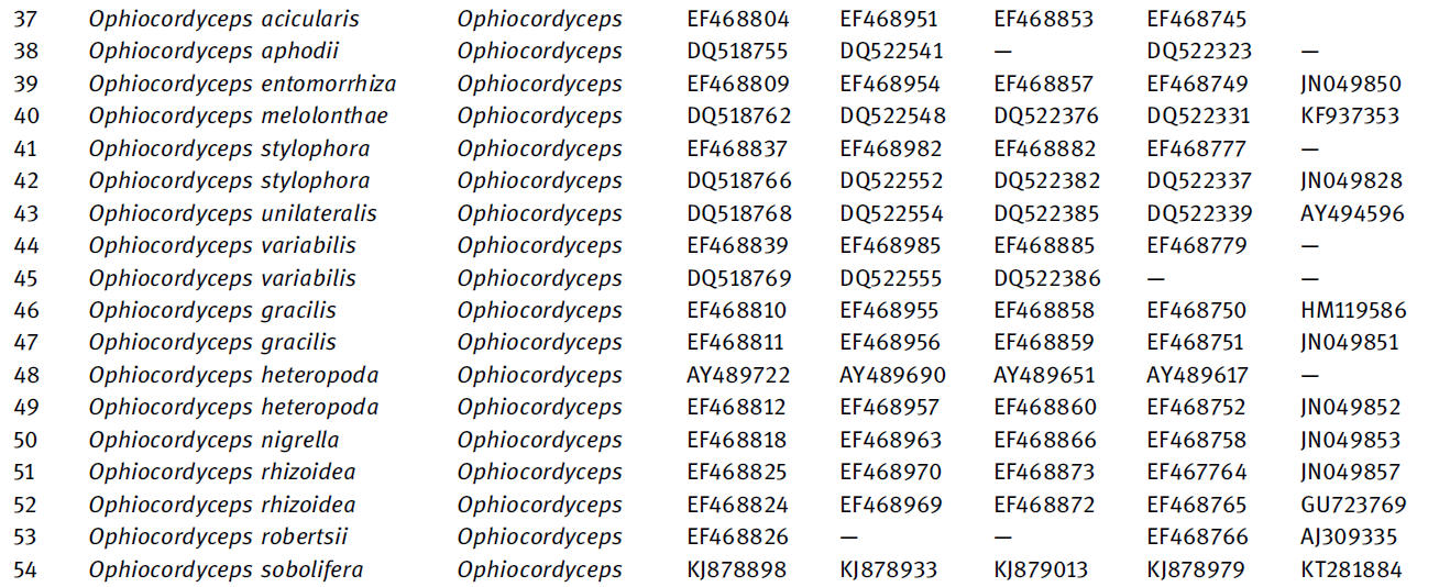
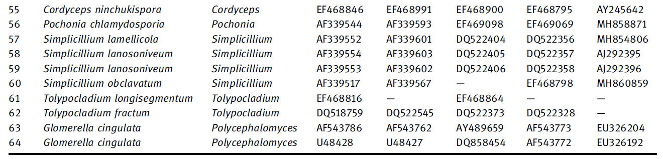
The data set of ITS, nrLSU, nrSSU, Rpb1, and Tef1 sequences were established by sequences downloaded from Genbank(NCBI) and based on the previous data published by Sung et al. (2007) [16]. The ITS, nrLSU, nrSSU, Rpb1, and Tef1 were noted with accession number, name of taxon, and locality.The multiple gene data used in the current study were established based on the combination of ITS, nrLSU, nrSSU,Rpb1, and Tef1 data. The amplified DNA sequences were proofread to remove ambiguous signals at both ends by different software, including Seaview 4.2.12 and Chromas Lite 2.1.1. The phylogenetic tree was constructed based on the neighbor-joining (NJ), maximum parsimony (MP), and maximum likelihood (ML) by using Molecular Evolutionary Genetics Analysis (MEGA) version X. Additionally, the best evolution model was predicted by using jModelTest.
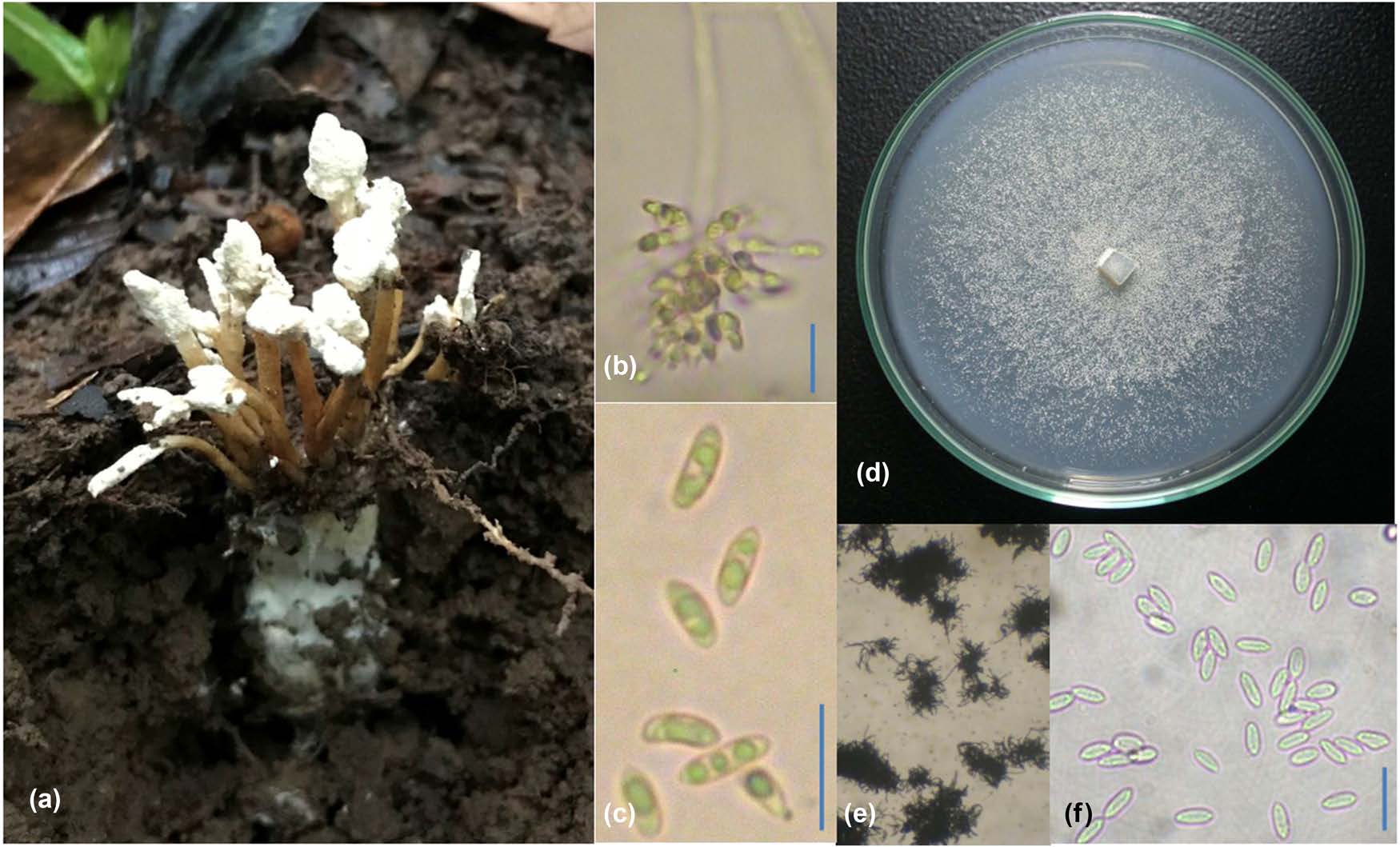
Figure 1: Morphology of Isaria cicadae: (a) Synemata forming from Cicadae, (b) Phialides, (c) conidia, (d) mycelia after 30 days on PDA media, (e) chain of conidia, and (f) conidia from mycelia. The bar scale indicated 10 μm.
3 Results
3.1 Morphology analysis
The sample of T011 was collected in the soil of the coffee garden on the morning of June 24, 2018. The synnemata were emerging from the soil, while the host was in the soil. Host: unidentified cicada nymph. Synnemata: presence,branching, 15–60mm in length × 1.0–2.5mm in diameter. Synnemata originated from the head of cicada nymphs with the thick layer of mycelia (hiding under the ground). Color: white to cream. Form: simple, erect, and distinguishing form fertile part and stipe. Fertile part: a dense white powdery covering on the surface due to the presence of a mass of conidia. Phialides: grouped inside the fertile part. Conidia: hyaline to white, cylindrical, 4.7–6.5 μm in length × 2.6–3.1 μm in diameter. Colonies from cultures on PDA: floccose and white, then becoming powdery by the conidiation of aerial hyphae. Conidia from the mycelia: smaller than those of from synnemata (Figure 1).
3.2 Amplification of ITS, nrLSU, nrSSU,Rpb1, and Tef1 gene
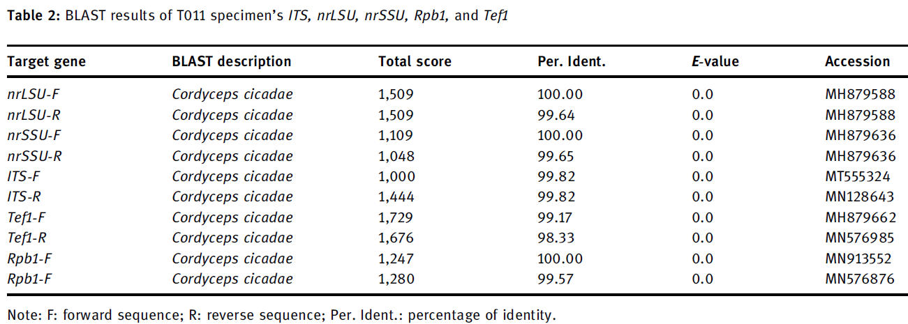
Isolated genomic DNA was amplified with the described primers; then, electrophoresis on 2.0% agarose gel showed a significant and clear band of gene ITS: 700 bps, nrLSU: 950 bps, nrSSU: 1,102bps, Rpb1: 803bps, and Tef1:1,020 bps. The PCR product was sequenced. Sequencing signals of both strands of both target genes were unique and good for reading (data not shown). According to BLAST results, the ITS, nrLSU, nrSSU, Rpb1, and Tef1 of T011 were similar to ITS, nrLSU, nrSSU, Rpb1, and Tef1 of C. cicadae (Telemorph of I. cicadae) (Table 2).