接第一篇
4. Taxonomy
4.1. Cordyceps cicadae (miq.) Massee, annals of botany 9: 38 (1895) [MB#311793]
4.1.1. Asexual morphs
Synnemata erect, irregularly branched, clavate, with powdery and floccose apex due to the conidial mass, white (2.5Y, 9.5/1) to pale yellow (2.5Y, 8.5/2) mycelium, 30–50mm in length and 0.5–2.5mm in diam (Figures 1(A) and 2(A)). Conidiophores and Phialides were not observed. Conidia narrowly cylindrical with round apex, sometimes slightly curved, smooth, thin-walled, hyaline, containing one or two guttules, 6.0–7.5×2.0–3.5 μm [Q=2.14–3.25,Q=2.7 ± 0.5] (n=40).
4.1.2. Specimen examined
Korea. Gwangju Metropolitan City, Mudeungsan National Park, 35°07'51ʺ N, 126°59'62ʺ E, mixed hardwood forest, parasitic in cicada, 24 Sep 2020, S.L. Kwon, KUC20200924-44 (NIBRFG0000511371).
4.1.3. Remarks
Cordyceps cicadae KUC20200924-44 matched well with C. cicadae (Miq.) Massee in the phylogenetic analysis. The size of the conidia was measured and found to be similar to the original description [55].Further microscopic observations are required for accurate identification. According to phylogenetic analysis, C. qingchengensis is the most closely related species. The difference between C. cicadae and C. qingchengensis is the host species. The hosts of C. cicadae are cicada nymphs, while the host of C. qingchengensis has been identified to be the cocooned pupa of a large silk moth [21]. However, the asexual morph of C. qingchengensis is unknown; therefore, it was impossible to compare its morphological characteristics.
4.2. Dentocorticium bicolor (P.H.B. Talbot)Nakasone & S.H. He, MycoKeys 32: 42(2018) [MB#823073]
Basidiocarp annual, resupinate, loosely adnate, effused, little tough, easily separated from substrate when dried, up to 200–300 μm thick (Figures 1(B) and 2(B)). Hymenial surface membranaceous, slightly odontoid, hydonoid, tuberculate because of hyphal pegs, margin abrupt, very pale yellow (2.5Y, 9.5/2) to yellow (5Y, 8/8) when fresh, pale yellow (5Y, 8/2) to yellow (5Y, 8/6) when dry. Hyphal system dimitic,generative hyphae located in subhymenium, moderately septate with clamp connections, branched, thinwalled, hyaline, 2.0–3.5 μm in diam. Skeletal hyphae easily separated from generative hyphae, long, straight, aseptate with clamp connections, unbranched, thickwalled up to 1 μm with thin-walled round apex, brownish yellow (10YR, 6/6), 2.7–4.1 μm in diam. Dendrohyphidia arising from subhymenial hyphae,irregularly and strongly branched, tortuous and with bulges, thin-walled, hyaline, 1.0–2.5 μm in diam.Basidia clavate, 4-spored, smooth, hyaline, containing one or two guttules (17–)18.5–21(–21.5) ×4.0–5.0 μm (n=31). Basidiospores ellipsoid to cylindrical,smooth, thin-walled, hyaline, containing over two guttules,7.5–8.5×3.0–4.5 μm [Q=1.9–2.5, Q=2.2 ± 0.3](n=34). Cystidia lacks, cylindrical to subclavate,smooth, thin-walled, hyaline, 17–27×3.7–6.2 μm(n=8).
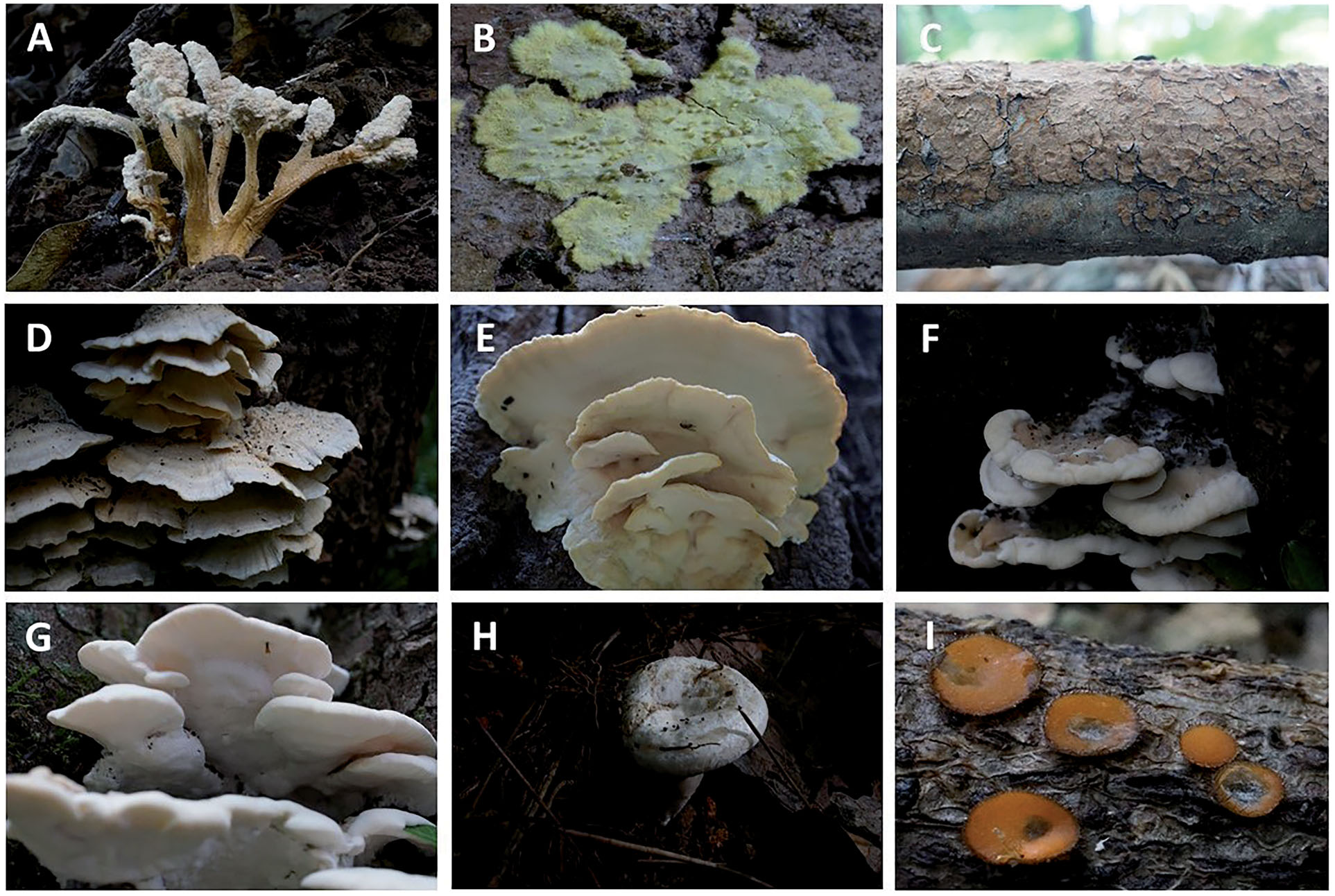
Figure 1. Fruiting bodies of the seven unrecorded species in Korea.
(A) Cordyceps cicadae;
(B) Dentocorticium bicolor;
(C) Hymenochaete nanospora;
(D and E) Physisporinus crataegi;
(F and G) Rigidoporus piceicola;
(H) Russula raoultii;
(I)Scutellinia crinita.
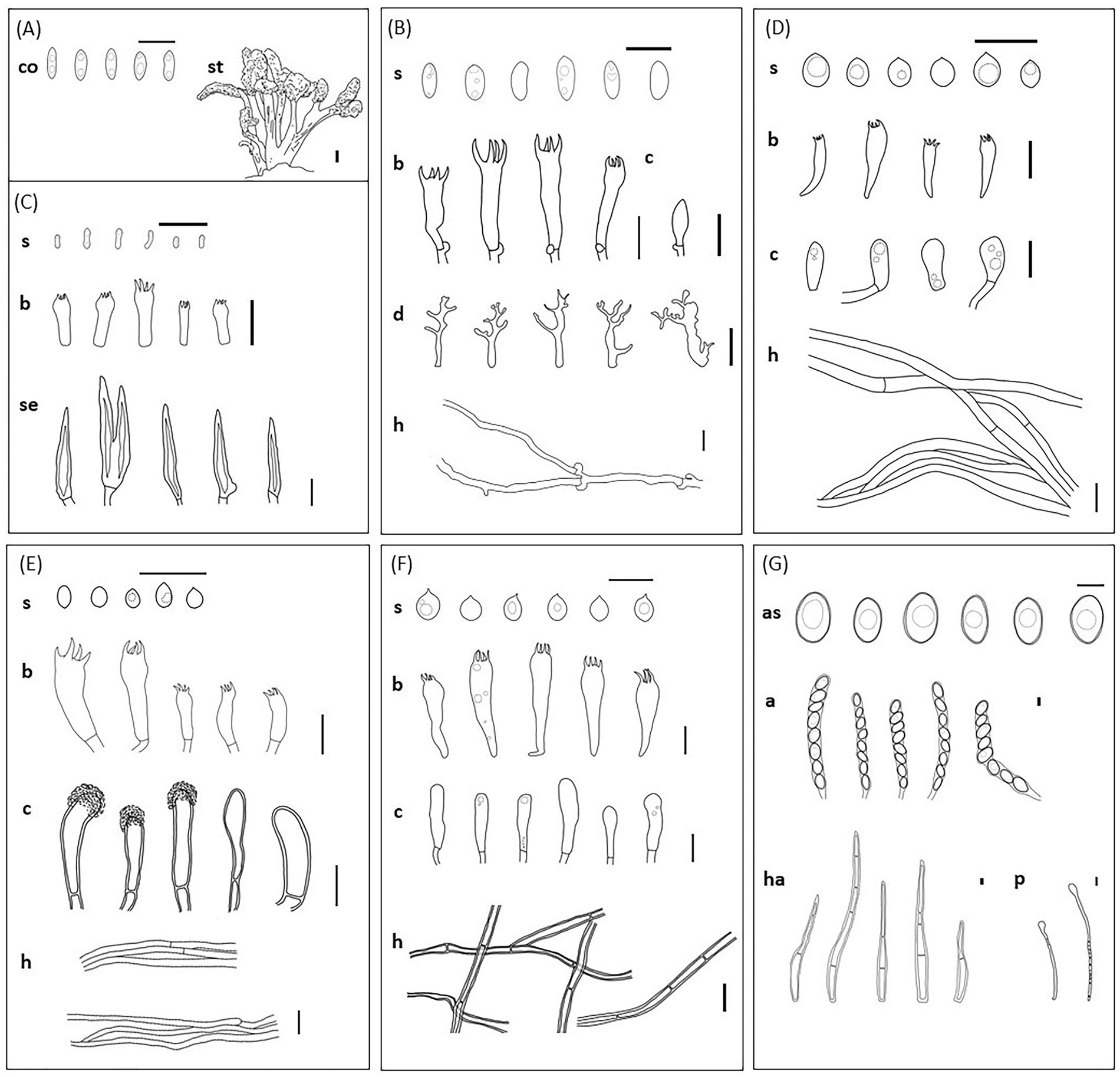
Figure 2. Microscopic view of morphological characteristics of the seven unrecorded species in Korea.
(A) Cordyceps cicadae;
(B) Dentocorticium bicolor;
(C) Hymenochaete nanospora;
(D) Physisporinus crataegi; (E) Rigidoporus piceicola; (F) Russula raoultii;
(G) Scutellinia crinita. (a: asci, as: ascospores, b: basidia, c: cystidia, co: conidia, d: dendrohyphidia, h: hyphae, ha: hair, p: paraphyses,s: basidiospores, se: setae, st: stroma; a scale bar=10 μm, for stroma, a scale bar=1 cm).
4.2.1. Specimen examined
Korea. Gwangju Metropolitan City, Mudeungsan National Park, 35°08'80ʺ N, 126°58'59ʺ E, mixed hardwood forest, occurring on dead trees, 27 May 2020, S. L. Kwon, KUC20200527-18(NIBRFG0000507813), 17 June 2020, S. L. Kwon,KUC20200617-44 (NIBRFG0000508874).
4.2.2. Remarks
Dentocorticium bicolor KUC20200527-18 and KUC20200617-44 matched well with D. bicolor (P.H.B. Talbot) Hjortstam & Ryvarden in the phylogenetic analysis, with high bootstrap value.The morphological characteristics of D. bicolor KUC20200527-18 and KUC20200617-44 are similar to the original description. According to the description of D. bicolor (Talbot) Hjortstam & Ryvarden, the size of the basidia was determined to be 30–35×6–7 μm. However, according to Grandinia bicolor (P.H.B. Talbot), a synonym of D.bicolor, the basidia size is smaller than that of D.bicolor, KUC20200527-18 and KUC20200617-44. In addition, the context color was reported as brown; however, brown-colored samples were not observed. Further studies are required to obtain more accurate results. Phylogenetic analysis shows that D. sulphurellum is the most closely related species. D. sulphurellum has white to yellow hymenial surface and basidiospores (7–9.5×2.5–3 μm), similar to D.bicolor. However, D. sulphurellum has only been reported in North America [30].
4.3. Hymenochaete nanospora J.C. Léger,
cryptogamie mycologie 4 (3): 235(1983) [MB#108064] Basidiocarps annual, resupinate, effused, adherent,margin thin, woody hard when dry, cortex present(Figures 1(C) and 2(C)). Hymenial surface crustaceous,smooth, densely cracked, light brown(7.5YR, 6/4) when fresh, brown (7.5YR, 4/4) when dry. Hyphal system monomitic, usually septate without clamp connections, occasionally branched,thin-walled, hyaline, 2.1–3.3 mm in diam. Setae obclavate, fusiform, with acute apex, partition absent, smooth, thick-walled, reddish brown (7.5YR,6/8), 20–45×4.0–5.0 μm (=30). Basidia clavate,subcylindrical, 4-spored, smooth, hyaline (8.5–)9.0–11.5(–12.5)×2.0–3.5 μm (n=48). Basidiospores narrowly cylindrical to allantoid, smooth, thinwalled,hyaline, containing two guttules, 2.5–3.5×0.8–1.2 μm [Q=2.92–3.125, Q=3.02 ± 0.1] (n=23).Cystidia absent.
4.3.1. Specimen examined
Korea. Gwangju Metropolitan City, Mudeungsan National Park, 35°07'52ʺ N, 126°59'62ʺ E, mixed hardwood forest, occurring on hardwood branches,24 Sep 2020, S. L. Kwon, KUC20200924-48 (NIBR FG0000511372).
4.3.2. Remarks
Hymenochaete nanospora KUC20200924-48 was well-matched with H. nanospora J.C. Léger in the phylogenetic analysis. This species is morphologically characterized by small and narrow spores, and its width rarely exceeds 1 μm. According to phylogenetic analysis, H. cana was identified as the most closely related species. H. cana has reddish brown setae (20–40×5.0–8.0 μm) like H. nanospora.However, H. cana differs from H. nanospora in that it has mouse-gray to ash-gray basidiocarps, without a cortex, and larger basidiospores (2.8–)3–3.4(–3.6)×(1.5–)1.6–1.9(–2) μm [56].
4.4. Physisporinus crataegi F. Wu, jia J. Chen &
Y.C. Dai, mycologia 109 (5): 751(2017) [MB#819191]Basidiocarps annual, effused-reflexed, pileate, mostly imbricate, soft when fresh, fragile when dry (Figures1(D,E) and 2(D)). Pileus elongnated, up to 4 cm long, 8 cm wide, and 4.5 cm thick at the base. Pileus surface zonate, margin crenate, incurved when dry, pale orange yellow (10YR, 9.5/2) to very pale brown (10YR, 8.5/2). Pores angular to round, 6–7 per mm,dissepiments thin, lacerate. Pore surface white (2.5Y,8/1) to yellow (2.5Y, 8/6) when fresh, becoming dark yellowish brown (18YR, 4/6) when dry. Context corky, azonate, 1.2mm thick. Tubes concolorous with surface of the pores, tube layers distinct, each layer up to 5mm long. Hyphal system monomitic, hyphae usually septate and occasionally with clamp connections, sometimes branched, thin-walled, hyaline,3.5–5.5 μm in diam. Basidia broadly clavate, 4-spored, smooth, hyaline (8.0–)10.5–13.5×4.5–5.5 mm(n=17). Basidiospores ellipsoid, smooth, thinwalled,hyaline, sometimes containing a guttle,4.5–5.0(–5.5)×3.0–4.0 μm [Q=1.25–1.5, Q=1.38 ±0.13] (n=44). Hymenial cystidia absent. Cystidioles present, clavate, ovoid, fusoid, smooth, thin-walled,hyaline, 10–14.5×4.5–6.5 μm (n=39).
4.4.1. Specimen examined
Korea. Gwangju Metropolitan City, Mudeungsan National Park, 35°08'59ʺ N, 126°58'97ʺ E, mixed hardwood forest, clustered or mesic in the lower part of a broad-leaved tree stump, 3 Sep 2020, S. L.Kwon, KUC20200903-15 (NIBRFG0000511374).
4.4.2. Remarks
Physisporinus crataegi KUC20200903-15 is matched well with P. crataegi F. Wu, Jia. J. Chen & Y.C. Dai,Wu, Chen, Ji, Vlasak & Dai in the phylogenetic analysis. Besides the presence of various cystidioles observed on the new species, microscopic characteristics of P. crataegi KUC20200903-15 match the original description [20]. P. crataegi is characterized by effused-reflexed and soft basidiocarps, cystidia absent, and ellipsoid basidiospores, and grows on broad-leaved trees in temperate forests. The resupinate region of P. crataegi is similar to that of P. vitreus in having soft basidiocarps, but P. vitreus has larger basidiospores (5–6×4–5 μm) and pores (4–6 per mm) [20]. Phylogenetically, P. cinereus was identified as the most closely related species (Núnez & Ryvarden) F. Wu et al. However, P. cinereus has fibrillose and grayish pileus, large pores (5–6 per mm), and large globose-shaped basidiospores, 5–6 mm in diam [57], whereas P. crataegi has glabrous and cream-colored pileus with distinct pellicle,smaller pores (6–8 per mm), and broadly ellipsoid to subglobose basidiospores (4.5–5.0(–5.5)×3.0–4.0 μm). In addition, the basidiocarp of P. crataegi is fragile when dry, whereas that of P. cinereus is woody hard [57].